Research Article | Volume 11, Issue 3, May, 2023

Effect of bacteriophages and chamber bitter (Phyllanthus amarus) in combination on Vibrio parahaemolyticus

Truong Thi Bich Van Do Tan Khang Le Viet Dung Le Hoang Bao Ngoc Nguyen Pham Anh Thi Tran Thanh Men Vo Ngoc Tram Anh Nguyen Kim Thoa   

Open Access   

Published:  Apr 04, 2023

DOI: 10.7324/JABB.2023.91082
Abstract

Vibrio parahaemolyticus is one of the causes of acute hepatopancreatic necrosis disease or early mortality syndrome in shrimps. Frequent and repeated application of antibiotics leads to generating resistant strains. Bacteriophages and plant extracts are considered potential biological agents in precaution and treatment of bacterial diseases in aquaculture. The study aimed to investigate the effects of bacteriophages and Phyllanthus amarus extract on strains of V. parahaemolyticus bacteria that were isolated from white leg shrimps (Litopenaeus vannamei). In this study, two strains of V. parahaemolyticus were used: B4XT4 (pure strain) and B4X0T2.2 (isolated strain) for preparing four treatments consisting of combining the bacteria with P. amarus extracts (8 mg/mL), bacteriophage (106 PFU/mL), a combination of P. amarus and bacteriophage, and a control treatment. The experiment was conducted in triplicate. The results showed that only plant extracts could inhibit bacterial growth. The colonial morphology of bacteria was changed when bacteriophages and P. amarus extract were added.


Keyword:     Bacteriophages Extract Inhibitory activity Phyllanthus amarus Vibrio parahaemolyticus


Citation:

Van TT, Khang DT, Dung LV, Ngoc LH, Thi NP, Men TP, Anh VN, Thoa NK. Effect of bacteriophages and chamber bitter (Phyllanthus amarus) in combination on Vibrio parahaemolyticus. J App Biol Biotech. 2023;11(3):70-76. https://doi.org/10.7324/JABB.2023.91082

Copyright: Author(s). This is an Open Access article distributed under the terms of the Creative Commons Attribution-NonCommercial-ShareAlike license.

HTML Full Text

1. INTRODUCTION

Vibrio parahaemolyticus has been one of the causes of acute hepatopancreatic necrosis disease or early mortality syndrome in shrimps in recent years. Using antibiotics or chemicals to inhibit bacteria has been the most popular course of treatment due to the initial benefits they bring. However, the long-term use of antibiotics leads to antibiotic resistance in bacteria, which is still a challenge in the aquaculture industry and the field of shrimp farming. The research on the inhibition of V. parahaemolyticus using biological therapy is being considered. The method of using phages against aquatic pathogenic bacteria was first used in Japan [1] and quickly became the subject of scientific interest. In recent years, there have been a number of studies using phages in the prevention and treatment of plant and animal diseases [2-4]. Besides, there are also many studies on plant extracts with high antimicrobial efficiency [5], especially Phyllanthus amarus [6,7]. Bacteriophages and P. amarus are considered potential biological agents in the treatment of bacterial diseases in aquatic animals. At present, research and potential applications of these agents in practice are underway, but studies in our country that involve their combined use are still limited. Therefore, the study of the ability of the phage in combination with the extract of this plant to inhibit the V. parahaemolyticus bacteria serves as a scientific basis for herbal applications in aquaculture in general and shrimp farming. The objective of this study was to investigate the interaction of phage and P. amarus extract and their effectiveness in inhibiting the spread of the pathogenic V. parahaemolyticus on white leg shrimp.


2. MATERIALS AND METHODS

2.1. Materials

All chemicals, equipment, and bacteriophages were supplied by the Molecular Biology Laboratory, Biotechnology Research and Development Institute, Cantho University. The methanolic extract of P. amarus (all the body) was obtained by the College of Natural Sciences, Cantho University. Bacteria strains were isolated from diseased shrimps, pond water, and mud in the Mekong Delta, Viet Nam.

2.2. Isolation of Vibrio spp.

Bacteria were isolated from diseased shrimps, pond water, and mud in the Vinh Thuan district, Kien Giang, Bac Lieu Provinces. Diseased shrimps were collected and dissected for hepatopancreas and intestines and were homogenized in a tryptic soy broth (TSB) medium supplemented with 1.5% NaCl and spread on a thiosulfate citrate bile salt sucrose (TCBS) medium. Water and mud samples were added to a TSB medium supplemented with 1.5% NaCl, incubated for 24 h, and spread on a TCBS medium. Green colonies were selected for further study.

2.3. Identification ofV. parahaemolyticus by PCR

V. parahaemolyticus was identified using two sets of primers. The amplification of the 16S rRNA gene with 700 bp amplicons using primer F (5’-CAGGCCTAACACATGCAAGTC-3’) and primer R (5’-GCATCTGAGTGTCAGTATCTGTCC-3’) was described by Mohamed et al. (2017) for identifying Vibrio spp. Strains that possessed the needed bands were then tested for their virulence properties using the ToxR gene F (5’- GTCTTCTGACGCAATCGTTG -3’) and R (5’- ATACGAGTGGTTGCTGTCATG -3’) which is specific for V. parahaemolyticus (Kim et al., 1999). The ToxR gene was amplified with 368 bp amplicons.

2.4. Double Layer Agar Technique

The test used the Double Agar Layer Technique. Two agar layers were prepared for 2 host tests: soft agar (0.3% agar, low agar concentration) was mixed with bacteria which was incubated in 24 h and overlaid on a hard agar layer (1.5% agar). The medium used in this study was King B supplemented with 0.5% NaCl. Afterward, 1 μL of bacteriophage was inoculated and incubated for 24 h to observe the plaques. The experiment was repeated once.

2.5. Plating Method

The experiment was conducted with four treatments where the extract and bacteriophage concentrations were used for testing, and they were 8 μg/mL and 20% of 106 PFU/mL, respectively, (treatment A consisted of 100 μL of bacteria; treatment B consisted of 100 μL of bacteria and 2 μL of extract; treatment C consisted of 100 μL of bacteria and 2 μL of bacteriophages; and treatment D consisted of 100 μL of bacteria, 2 μL of extract and 2 μL of bacteriophages).

The number and the morphology of colonies were recorded after 1 h, 3 h, and 24 h. The experiment was replicated 3 times, and the Stagraphic Centurion XVIII was used to analyze the data of a number of colonies. The size of colonies was observed using the CountPHICS software.


3. RESULTS

3.1. Isolation and Identification of V. parahaemolyticus

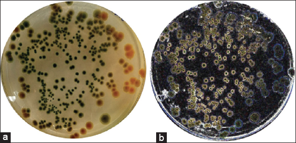
There were 48 bacterial strains that could grow on the TCBS media. All strains of bacteria were isolated from diseased shrimp with a morphological characterization similar to that of Vibrio spp. The colonies of these bacteria appeared circular, convex, and glossy. They were green in color and exhibited a diameter of about 1–2 mm on the TCBS medium after 24 h of incubation [8-10] [Figure 1]. For further confirmation, all the isolates were screened using 16S rRNA [Figure 2] and ToxR [Figure 3] primers, and were at 700 bp and 368 bp, respectively [11]. This study isolated 45 strains of Vibrio sp., including 8 strains of V. parahaemolyticus from diseased shrimp, pond water, and mud. B4XT4 and B4X0T2.2 strains were selected for the following experiments.

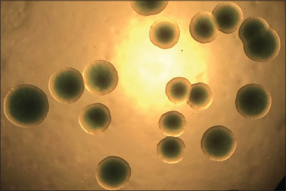
Figure 1: Vibrio parahaemolyticus colony morphology on TCBS agar.


[Click here to view]
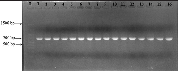
Figure 2: Gel electrophoresis of PCR products of 16S rRNA gene. (L: 1500bp ladder; 1: B4X0T2.1; 2: B4X0T2.2; 3: B4X0T2.3; 4: V4.1; 5: B4XCT1.1; 6: B4XCT1.2; 7: V4.2; 8: B4XCT1.2; 9: B4XCT1.5; 10: V4.3; 11: B3XD2; 12: V4.6; 13: B3DX4; 14: V4.7; 15: B3XD1; 16: V4.10).


[Click here to view]
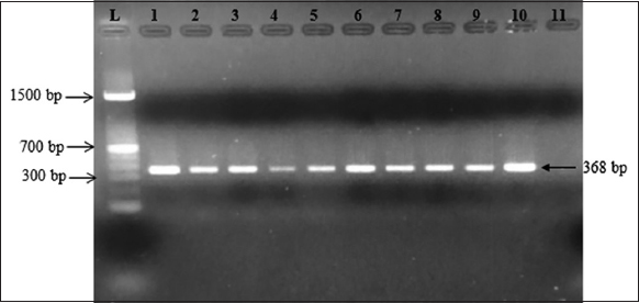
Figure 3: Gel electrophoresis of PCR products of toxR gene (L: 1500 bp ladder; 1: positive control; 2: B4X0T2.2; 3: B4X0T2.3; 4: B3XD2; 5: V4.1; 6: V4.2; 7: V4.3; 8: V4.6; 9: V4.6; 10: V4.10; 11: negative control).


[Click here to view]

3.2. Evaluation of Host Range

The infectiveness of 5 bacteriophages against 2 strains of V. parahaemolyticus B4XT4 and V. parahaemolyticus B4X0T2.2 is noted in Table 1. It is noteworthy that phage VD3 and phage VD4 displayed the possibility of infecting both strains of V. parahaemolyticus, but strains phage VD1 and phage VD5 only infected V. parahaemolyticus B4X0T2.2. On the other hand, the strain phage VD2 could infect neither V. parahaemolyticus B4XT4 nor V. parahaemolyticus B4X0T2.2.

Table 1: Evaluation of inhibitory activity on V. parahaemolyticus of bacteriophages.

Vibrio parahaelyticusPhage VD1Phage VD2Phage VD3Phage VD4Phage VD5
B4XT4*--++-
B4X0T2.2+-+++

+: Showed the phages capable of infecting host bacteria, - sign indicates that the phages incapable of infecting host bacteria.

3.3. Number of Colony Formation

The ability of the extract and phage to inhibit the growth of V. parahaemolyticus B4XT4 and V. parahaemolyticus B4X0T2.2 was different [Figure 4] after four treatments incubated at 1 h and 3 h. The number of colonies when adding extracts or phages both increased the number of bacteria compared to certificates. For V. parahaemolyticus B4XT4, when combining the phage and extract, the inhibitory effect on bacterial growth was more obvious with only the extract (the value decreased from 7.71 to 7.21 Log CFU/mL). For V. parahaemolyticus B4X0T2.2, it was the opposite, as adding the extract activated its growth. The study which was combined phage and P. amarus extract to inhibit the growth of V. parahaemolytiucs; however, initially there was no effect.

Figure 4: Interaction between extract and phage on bacterial population (a) affecting Vibrio parahaemolyticus B4XT4. (b) Affecting Vibrio parahaemolyticus B4X0T2.2.


[Click here to view]

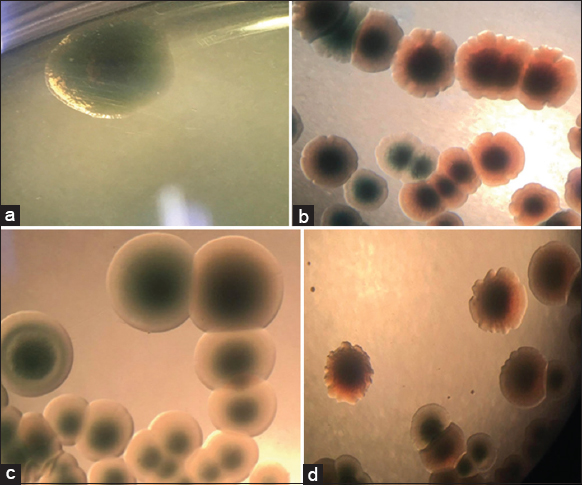
3.4. Effect of Plant Extract on the Colony of V. parahaemolyticus

Both strains of V. parahaemolyticus (B4XT4 and B4X0T2.2) were affected by the extract P. amarus, which changed the colony’s phenotype. The extract affected the shape of colonies — it caused the edge of V. parahaemolyticus B4X0T2.2 shrink and dentate with sharp and small peaks, compared to the round colonies of the control [Figure 5a and b]. The cover of the colonies was toothed, and lobate margins arose for B4XT4 [Figure 5c and d]. With regard to the colonies of V. parahaemolyticus B4X0T2.2, the size is smaller but insignificant. The most significant change in colony size was observed during a sharp decrease of 4 mm2 and 4.5 mm2, which was a drop of 1.3 times and 3.0 times, respectively [Figure 6a and b]. The decline in colony size apparently occurred in V. parahaemolyticus B4XT4; indeed, a size of 6 mm2 và 6.5 mm2 is notable, with a drop of 1.2 times and 1.1 times, respectively [Figure 6c and d].

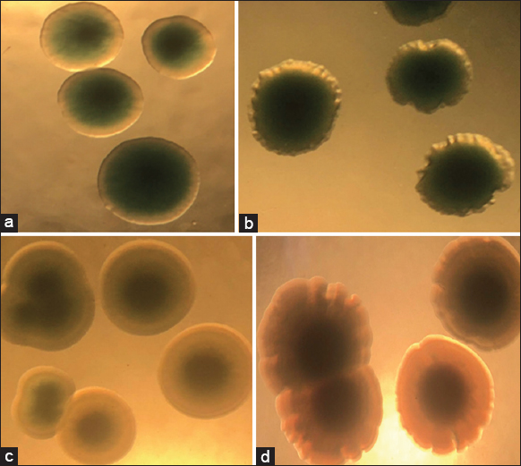
Figure 5: Effect of plant extract on colonies of Vibrio parahaemolyticus B4XT4 and B4X0T2.2. (a) B4XT4; (b) B4XT4+ plant extract; (c) B4X0T2.2; (d) B4X0T2.2+ plant extract.


[Click here to view]
Figure 6: Colony size changes of Vibrio parahaemolyticus B4X0T2.2 and B4XT4 in the presence of Phyllanthus amarus extract. (a) B4X0T2.2; (b) B4X0T2.2+ plant extract; (c) B4XT4; (d) B4XT4+ plant extract.


[Click here to view]

3.5. Changes in Colonial Morphology in the Presence of Bacteriophages

The colonial morphology of V. parahaaemolyticus B4XT4 and V. parahaaemolyticus B4X0T2.2, when infected with a phage, changed compared with the control. PhageVD1 infected V. parahaemolyticus B4XT4 and V. parahaaemolyticus B4X0T2.2, causing the colony to shrink and change its serration compared to the control [Figure 7].

Figure 7: VD1 changes colony phenotype and size of Vibrio parahaemolyticus B4X0T2.2 and B4XT4. (a) B4X0T2.2; (b) B4X0T2.2+phageVD3; (c) B4XT4; (d) B4XT4+phageVD1.


[Click here to view]

3.6. Impact of Plant Extract and Phages on V. parahaemolyticus

A comparison of the colony sizes in treatment between phage VD1, that could infect both strains V. parahaemolyticus, and phage VD3, which could only infect B4XT4, was conducted. As expected, the colony sizes of the former experienced a remarkable decline [Figure 8], though the latter made V. parahaemolyticus B4X0T2.2 remain the size of the colony [Figure 9].

Figure 8: Colony size changes Vibrio parahaemolyticus B4X0T2.2 and B4XT4 in the combination of Phyllanthus amarus extract and bacteriophage. (a) B4X0T2.2; (b) B4X0T2.2+ plant extract+phageVD1; (c) B4XT4; (d) B4XT4+ plant extract+phageVD1.


[Click here to view]
Figure 9: Colony size changes Vibrio parahaemolyticus B4X0T2.2 and B4XT4 in the combination of Phyllanthus amarus extract and bacteriophage. (a) B4X0T2.2; (b) B4X0T2.2+ plant extract+phageVD3; (c) B4XT4; and (d) B4XT4+ plant extract+phageVD3.


[Click here to view]

4. DISCUSSION

There have been some studies utilizing bacteriophages and P. amarus to inhibit V. parahaemolyticus but it was combined in single. Even so, all those factors presented their antimicrobacterial ability [12,13]. Unfortunately, the plant extract did not show an inhibitory effect in this experiment since the number of colonies still remained. That is possibly due to the fact that the concentration and incubation time of the extract was not optimal, so we could not conclude the efficacy of the plant extract in this regard. In addition, most of the experiments involving the use of plant extracts or bacteriophages combined with plant extracts to inhibit bacteria utilized the agar plate diffusion method to test the susceptibility of bacteria. However, this method has certain limitations — for example, molecules with a higher molecular weight diffuse very slowly in the agar, making it impossible to accurately measure the diameter of the halo ring in the plate. In contrast, in this experiment, the change of colonies was observed by using the plate count method. Although the effect of the extract on V. parahaemolyticus has not been elucidated, the colony form changed as they were affected by bacteriophages and the extracts clearly recorded the same.

Some studies on the topic of bacteriophages combined with plant extracts showed that the results were not satisfactory, except for the separate combination of the two factors [14]. Which used two types of bacteriophages (which have been identified) and three herbs combined to inhibit E. coli and concluded that it is necessary to further investigate the concentration of extracts and phages as well as combine many other ingredients to ensure the best results. The phage concentrations in this study were considered low because they were diluted to 5 times less than the original concentration. That prompted the use of the compound carvacrol, which is a compound found in many herbal essential oils (marjoram essential oil, essential oil of lemon basil, etc.), to achieve a purity rate of 99%. This only acted on but not completely inhibits the spread of the bacteria. In the experiment of [15] it was also found that the number of bacteria increased over time in the presence of bacteriophages; whereas the addition of carvacrol prevented the bacteria from becoming resistant to phages. Another critical point to note is that carvacrol is a purified substance, whereas in this experiment, we used P. amarus extract, which contains many unknown compounds that could work against the target bacteria.

Incidentally, it can be observed that there are some differences in colony morphology when treated with phages and P. amarus extract. The colony shapes were not round, but were spread out like a propeller, and such a colony form was referred to as a “competition type” [16]. That meant that the bacteria were motivated against the inhibitory factors that led to formation of the competition type. It is predicted that there existed a hidden genetic variation, which caused new phenotypes, and it was proved by True and Lindquist [17] on a yeast prion. Therefore, this experiment aimed to limit the pathogenicity of V. parahaemolyticus by making their genome not as intact as before.

In addition, a unique feature of this study is the use of the countPHICS software to measure the diameter of bacterial colonies. This software was introduced by Brzozowska et al. [18] and was supposed to be more reliable than manual measurement. The μ number in the images was higher than normal, indicating that the image resolution was low. This was due to the fact that the photos on the discs were taken only at 8 MP resolution. Fortunately, the included image processing software (ImageJ) was able to correctly evaluate the colonies for processing and for retrieving colony size data [Figure 10]. The count PHICS software was highly recommended in the colony size measurement experiments.

Figure 10: Images of Vibrio parahaemolyticus B4X0T2.2 before and after undertaking the ImageJ software. (a) Vibrio parahaemolyticus B4X0T2.2 before undertaking the ImageJ software. (b) Vibrio parahaemolyticus B4X0T2.2 after undertaking the ImageJ software.


[Click here to view]

5. ACKNOWLEDGMENTS

The authors would like to thank the Department of Research Affairs, Institute of Food and Biotechnology, Can Tho University for their valuable support. This study is fully funded by the Ministry of Education and Training.


6. AUTHORS’ CONTRIBUTIONS

All authors made substantial contributions to conception and design, acquisition of data, or analysis and interpretation of data; took part in drafting the article or revising it critically for important intellectual content; agreed to submit to the current journal; gave final approval of the version to be published; and agreed to be accountable for all aspects of the work. All the authors are eligible to be an author as per the International Committee of Medical Journal Editors (ICMJE) requirements/guidelines.


7. FUNDING

The study was funded by the Ministry of Education and Training, Vietnam (Project code: B2020-TCT-03).


8. CONFLICTS OF INTEREST

The authors report no financial or any other conflicts of interest in this work.


9. ETHICAL APPROVALS

This study does not involve experiments on animals or human subjects


10. DATA AVAILABILITY

All data generated and analyzed are included within this research article.


11. PUBLISHER’S NOTE

This journal remains neutral with regard to jurisdictional claims in published institutional affiliation.

REFERENCES

1.  Nakai T, Sugimoto R, Park KH, Matsuoka S, Mori K, Nishioka T, et al. Protective effects of bacteriophage on experimental Lactococcus garvieae infection in yellowtail. Dis Aquat Organ 1999;37:33-41. [CrossRef]

2.  Tanji Y, Shimada T, Fukudomi H, Miyanaga K, Nakai Y, Unno H. Therapeutic use of phage cocktail for controlling Escherichia coli O157:H7 in gastrointestinal tract of mice. J Biosci Bioeng 2005;100:280-7. [CrossRef]

3.  Chen L, Fan J, Yan T, Liu Q, Yuan S, Zhang H, et al. Isolation and characterization of specific phages to prepare a cocktail preventing Vibrio sp. Va-F3 infections in shrimp (Litopenaeus vannamei). Front Microbiol 2019;10:2337. [CrossRef]

4.  Holtappels D, Fortuna K, Lavigne R, Wagemans J. The future of phage biocontrol in integrated plant protection for sustainable crop production. Curr Opin Biotechnol 2021;68:60-71. [CrossRef]

5.  Gonelimali FD, Lin J, Miao W, Xuan J, Charles F, Chen M, et al. Antimicrobial properties and mechanism of action of some plant extracts against food pathogens and spoilage microorganisms. Front Microbiol 2018;9:1639. [CrossRef]

6.  Ismail CA, Deris ZZ, Bakar RA, Ismail N. In vitro anti-leptospiral activity of Phyllanthus amarus extracts and their combinations with antibiotics. Int J Environ Res Public Health 2021;18:2834. [CrossRef]

7.  Paria P, Kunal SP, Behera BK, Mohapatra PK, Das A, Parida PK, et al. Molecular characterization and genetic diversity study of Vibrio parahaemolyticus isolated from aquaculture farms in India. Aquaculture 2019;509:104-11. [CrossRef]

8.  Hikmawati F, Susilowati A, Setyaningsih R. Colony morphology and molecular identification of Vibrio spp. on green mussels (Perna viridis) in Yogyakarta, Indonesia tourism beach areas. Biodivers J Biol Divers 2019;20:2891-9. [CrossRef]

9.  Tinh TH, Elayaraja S, Mabrok M, Gallantiswara PC, Vuddhakul V, Rodkhum C. Antibacterial spectrum of synthetic herbal-based polyphenols against Vibrio parahaemolyticus isolated from diseased pacific whiteleg shrimp (Penaeus vannamei) in Thailand. Aquaculture 2021;533:736070. [CrossRef]

10.  Zhang X, Sun J, Chen F, Qi H, Chen L, Sung YY, et al. Phenotypic and genomic characterization of a Vibrio parahaemolyticus strain causing disease in Penaeus vannamei provides insights into its niche adaptation and pathogenic mechanism. Microb Genom 2021;7:000549. [CrossRef]

11.  Ribeiro AM, de Sousa JN, Costa LM, Oliveira FA, Santos RC, et al. Antimicrobial activity of Phyllanthus amarus schumach. &Thonn and inhibition of the NorA efflux pump of Staphylococcus aureus by phyllanthin. Microb Pathog 2019;130:242-6. [CrossRef]

12.  Jun JW, Shin TH, Kim JH, Shin SP, Han JE, Heo GJ, et al. Bacteriophage therapy of a Vibrio parahaemolyticus infection caused by a multiple-antibiotic-resistant O3:K6 pandemic clinical strain. J Infect Dis 2014;210:72-8. [CrossRef]

13.  Phuong TV, Yen PT, Linh NQ. Antibacterial activity of extracts from dried and fresh herbal plant (Phyllanthus amarus) against pathogens causing acute hepatopancreatic necrosis disease (Ahpnd) in white leg shrimp (Litopenaeus vannamei) at thua thien hue province, Vietnam. Asp J Biomed Clin Case Rep 2019;2:120-8. [CrossRef]

14.  Pimchan T, Cooper CJ, Eumkeb G, Nilsson AS. In vitro activity of a combination of bacteriophages and antimicrobial plant extracts. Lett Appl Microbiol 2018;66:182-7. [CrossRef]

15.  Ni P, Wang L, Deng B, Jiu S, Ma C, Zhang C, et al. Combined application of bacteriophages and carvacrol in the control of Pseudomonas syringae pv. actinidiae planktonic and biofilm forms. Microorganisms 2020;8:837. [CrossRef]

16.  Blandchard AE, Lu T. Bacterial social interactions drive the emergence of differential spatial colony structure. BMC Syst Biol 2015;9:59. [CrossRef]

17.  True HL, Lindquist SL. A yeast prion provides a mechanism for genetic variation and phenotypic diversity. Nature 2000;407:477-83. [CrossRef]

18.  Brzozowska B, Ga?ecki M, Tartas A, Ginter J, Ka?mierczak U, Lundholm L. Freeware tool for analysing numbers and sizes of cell colonies. Radiat Environ Biophys 2019;58:109-17. https://doi.org/10.1007/s00411-018-00772-z PMid:30673853 PMCid:PMC6394662

Reference

1. Nakai T, Sugimoto R, Park KH, Matsuoka S, Mori K, Nishioka T, et al. Protective effects of bacteriophage on experimental Lactococcus garvieae infection in yellowtail. Dis Aquat Organ 1999;37:33-41. https://doi.org/10.3354/dao037033

2. Tanji Y, Shimada T, Fukudomi H, Miyanaga K, Nakai Y, Unno H. Therapeutic use of phage cocktail for controlling Escherichia coli O157:H7 in gastrointestinal tract of mice. J Biosci Bioeng 2005;100:280-7. https://doi.org/10.1263/jbb.100.280

3. Chen L, Fan J, Yan T, Liu Q, Yuan S, Zhang H, et al. Isolation and characterization of specific phages to prepare a cocktail preventing Vibrio sp. Va-F3 infections in shrimp (Litopenaeus vannamei). Front Microbiol 2019;10:2337. https://doi.org/10.3389/fmicb.2019.02337

4. Holtappels D, Fortuna K, Lavigne R, Wagemans J. The future of phage biocontrol in integrated plant protection for sustainable crop production. Curr Opin Biotechnol 2021;68:60-71. https://doi.org/10.1016/j.copbio.2020.08.016

5. Gonelimali FD, Lin J, Miao W, Xuan J, Charles F, Chen M, et al. Antimicrobial properties and mechanism of action of some plant extracts against food pathogens and spoilage microorganisms. Front Microbiol 2018;9:1639. https://doi.org/10.3389/fmicb.2018.01639

6. Ismail CA, Deris ZZ, Bakar RA, Ismail N. In vitro anti-leptospiral activity of Phyllanthus amarus extracts and their combinations with antibiotics. Int J Environ Res Public Health 2021;18:2834. https://doi.org/10.3390/ijerph18062834

7. Paria P, Kunal SP, Behera BK, Mohapatra PK, Das A, Parida PK, et al. Molecular characterization and genetic diversity study of Vibrio parahaemolyticus isolated from aquaculture farms in India. Aquaculture 2019;509:104-11. https://doi.org/10.1016/j.aquaculture.2019.04.076

8. Hikmawati F, Susilowati A, Setyaningsih R. Colony morphology and molecular identification of Vibrio spp. on green mussels (Perna viridis) in Yogyakarta, Indonesia tourism beach areas. Biodivers J Biol Divers 2019;20:2891-9. https://doi.org/10.13057/biodiv/d201015

9. Tinh TH, Elayaraja S, Mabrok M, Gallantiswara PC, Vuddhakul V, Rodkhum C. Antibacterial spectrum of synthetic herbal-based polyphenols against Vibrio parahaemolyticus isolated from diseased pacific whiteleg shrimp (Penaeus vannamei) in Thailand. Aquaculture 2021;533:736070. https://doi.org/10.1016/j.aquaculture.2020.736070

10. Zhang X, Sun J, Chen F, Qi H, Chen L, Sung YY, et al. Phenotypic and genomic characterization of a Vibrio parahaemolyticus strain causing disease in Penaeus vannamei provides insights into its niche adaptation and pathogenic mechanism. Microb Genom 2021;7:000549. https://doi.org/10.1099/mgen.0.000549

11. Ribeiro AM, de Sousa JN, Costa LM, Oliveira FA, Santos RC, et al. Antimicrobial activity of Phyllanthus amarus schumach. & Thonn and inhibition of the NorA efflux pump of Staphylococcus aureus by phyllanthin. Microb Pathog 2019;130:242-6. https://doi.org/10.1016/j.micpath.2019.03.012

12. Jun JW, Shin TH, Kim JH, Shin SP, Han JE, Heo GJ, et al. Bacteriophage therapy of a Vibrio parahaemolyticus infection caused by a multiple-antibiotic-resistant O3:K6 pandemic clinical strain. J Infect Dis 2014;210:72-8. https://doi.org/10.1093/infdis/jiu059

13. Phuong TV, Yen PT, Linh NQ. Antibacterial activity of extracts from dried and fresh herbal plant (Phyllanthus amarus) against pathogens causing acute hepatopancreatic necrosis disease (Ahpnd) in white leg shrimp (Litopenaeus vannamei) at thua thien hue province, Vietnam. Asp J Biomed Clin Case Rep 2019;2:120-8. https://doi.org/10.36502/2019/ASJBCCR.6173

14. Pimchan T, Cooper CJ, Eumkeb G, Nilsson AS. In vitro activity of a combination of bacteriophages and antimicrobial plant extracts. Lett Appl Microbiol 2018;66:182-7. https://doi.org/10.1111/lam.12838

15. Ni P, Wang L, Deng B, Jiu S, Ma C, Zhang C, et al. Combined application of bacteriophages and carvacrol in the control of Pseudomonas syringae pv. actinidiae planktonic and biofilm forms. Microorganisms 2020;8:837. https://doi.org/10.3390/microorganisms8060837

16. Blandchard AE, Lu T. Bacterial social interactions drive the emergence of differential spatial colony structure. BMC Syst Biol 2015;9:59. https://doi.org/10.1186/s12918-015-0188-5

17. True HL, Lindquist SL. A yeast prion provides a mechanism for genetic variation and phenotypic diversity. Nature 2000;407:477-83. https://doi.org/10.1038/35035005

18. Brzozowska B, Ga?ecki M, Tartas A, Ginter J, Ka?mierczak U, Lundholm L. Freeware tool for analysing numbers and sizes of cell colonies. Radiat Environ Biophys 2019;58:109-17. https://doi.org/10.1007/s00411-018-00772-z

Article Metrics
267 Views 167 Downloads 434 Total

Year

Month

Related Search

By author names

Similar Articles

A Brief Survey of Fungi as Allergen in Respiratory Allergic Patients by Intradermal Skin Sensitivity Test in Terai Area

Rachna Chaturvedi and Sushil Chaturvedi

Bioactivity assessment of endophytic fungi associated with Centella asiatica and Murraya koengii

Archana Nath, Jyoti Pathak and SR Joshi

Anti-candidal activities of some Myrtus communis L. extracts obtained using accelerated solvent extraction (ASE)

Elif Ayse Erdogan, Gulden Goksen, Ayse Everest

In vitro Antimicrobial Comparison of Taif and Egyptian Pomegranate Peels and Seeds Extracts

Ahmed Gaber , Mohamed M. Hassan , El-Dessoky S. Dessoky , Attia O. Attia

Antimicrobial Activity Screening of Marine Bacteria Isolated from the Machilipatnam Sea Coast of Andhra Pradesh, India

K. Bala Chandra, V. Umamaheswara Rao, Subhaswaraj Pattnaik, Siddhardha Busi

Effects of Vitex doniana leaf extract on aluminium-induced toxicity in male albino Wistar rats

O. E. Yakubu , O. F. C. Nwodo, C. Imo, M. Abdulrahaman, L. B. Uyeh

In Vitro Antidiabetic and Anti-oxidant Activities of Methanol Extract of Tinospora Sinensis

Anindita Banerjee, Bithin Maji, Sandip Mukherjee, Kausik Chaudhuri, Tapan Seal

Mild Acid Hydrolysis-related Release of Water-soluble Sunscreen Pigments from the Exopolysaccharide Matrix of Edible Terrestrial Cyanobacteria

Wen Liu, Haiyan Xu, Xiang Gao

Preventive and curative control of sclerotium rot disease of cocoyam cormel (Colocasia esculenta [L., Scott]) using plant extracts and Trichoderma koningii

Nwauzoma Akagbuo Bartholomew, Jaja Tamunodiari Emylia, Njoku Chibuzor

Comparative analyses of genomic DNA extracted from freshwater fish tissues preserved in formaldehyde and alcohol in different periods of time

R. K. Garg, Khushboo Sengar, R. K. Singh

Hepatoprotective activity of methanolic shoot extract of Bambusa bambos against carbon tetrachloride induce acute liver toxicity in Wistar rats

A. A. Shetti Suman Patil, H Joy Hoskeri, P Rajeev, Bhushan Kulkarni, Geetanjali R Kamble, Gurusiddhesh B Hiremath, Vishal Kalebar, S V Hiremath

Phytoextraction of Heavy Metals and Risk Associated with Vegetables Grown from Soil Irrigated with Refinery Wastewater

A. Y. Ugya, A. M. Ahmad, H. I. Adamu, S. M. Giwa, T. S. Imam

Polyphenols and triterpenes in leaves and extracts from three Nicotiana species

Venelina Popova, Tanya Ivanova, Albena Stoyanova, Violeta Nikolova, Tsveta Hristeva, Margarita Docheva, Nikolay Nikolov, Ivan Iliev

Antiproliferative activities of Althaea ludwigii L. extract on Michigan Cancer Foundation-7 breast cancer cell line

Zinah Essam Hameed Alshaya, Enas Jawad Kadhim, Hayder B. Sahib

Antibacterial activity of leaf extract of Chromolaena odorata and the effect of its combination with some conventional antibiotics on Pseudomonas aeruginosa isolated from wounds

P. Odinakachukwu Omeke, J. Okechukwu Obi, N. A. Ibuchukwu Orabueze , Anthony Chibuogwu Ike

Enhanced biobutanol production by optimization of the medium from Clostridium acetobutylicum MTCC 11274 using macroalgae Gracilaria edulis

Shelly Rana, Kashyap Dubey, Ashwani Dhingra

In vitro antiproliferative effect of aqueous extract of Solanum macranthum fruits on MDA-MB-231 tripple negative breast cancer cell line

Vishal U. Kalebar , Joy H. Hoskeri, Shivaprakash V. Hiremath, Murigendra B. Hiremath

Screening of in vitro sun protection factor of some medicinal plant extracts by ultraviolet spectroscopy method

Manisha Pralhad Sutar, Sanjay Ravindra Chaudhari

Antimicrobial activity of the lichens Parmotrema andium and Dirinaria applanata

Bijayananda Sahoo, Satyabrata Dash, Sabyasachy Parida, Jayanta Kumar Sahu, Biswajit Rath

Application of guava leaves extract on jelly candy to inhibit Streptococcus mutans

Yuniwaty Halim, Raphael Dimas Tri Nugroho, Hardoko,, Ratna Handayani

Effect of extraction techniques on anthocyanin from butterfly pea flowers (Clitoria ternatea L.) cultivated in Vietnam

Nguyen Minh Thuy, Tran Chi Ben, Vo Quang Minh, Ngo Van Tai

Interaction efficiency of Trichoderma spp. and some plant extracts against ear-cockle disease

Azher Hameed Faraj Al-Taie, Noor Kadhum Al-Zubaid

Optimization of extraction conditions of phytochemical compounds in “Xiem” banana peel powder using response surface methodology

Ngo Van Tai, Mai Nhat Linh, Nguyen Minh Thuy

Allelopathic effect of marigold (Tagetes erecta L.) leaf extract on growth of Chlorella vulgaris

Tassnapa Wongsnansilp, Wikit Phinrub, Niran Juntawong

Anti-proliferative activities of solasodine extracts from different Solanum spp. cell cultures on colon and bone carcinoma cell lines

Vijaykumar Deshmukh,, Sangeeta Ballav, Soumya Basu, Sanjay Mishra,, Jyoti Deshpande, Minal Wani

Pseudomonas gessardii—A novel pathogenic bacterium associated with the cases of corneal ulcers and producing virulent pyoverdine pigment

Deepika Jain

Growth-promoting effects of marine microalgae species using tropical forest soil extracts

Kasturi Arumugam, Nor Suhaila Yaacob, Hasdianty Abdullah, Mohd Fadzli Ahmad, Maegala Nallapan Maniyam, Emi Fazlina Hashim, Fridelina Sjahrir, Wan Muhammad Ikram, Kazuhiro Komatsu, Victor S. Kuwahara

Effect of extraction methods and temperature preservation on total anthocyanins compounds of Peristrophe bivalvis L. Merr leaf

Nguyen Minh Thuy, Dao Huynh Ngoc Han, Vo Quang Minh, Ngo Van Tai

Investigating Musa paradisiaca (Var. Nanjangud rasa bale) pseudostem in preventing hyperglycemia along with improvement of diabetic complications

Ramith Ramu,, Prithvi S Shirahatti, Deepika TH, Shrisha Naik Bajpe, Navya Sreepathi,, Shashank M Patil, Nagendra Prasad MN

Extraction and quantification of acrylic acid from acrylamidase-catalyzed reaction produced by Bacillus tequilensis

Riddhi Prabha, Vinod Kumar Nigam

Microplastics accumulation in agricultural soil: Evidence for the presence, potential effects, extraction, and current bioremediation approaches

Varsha Yadav, Saveena Dhanger, Jaigopal Sharma

Application of butterfly pea flower extract in processing some Vietnamese traditional foods

Nguyen Minh Thuy, Tran Chi Ben, Pham Thi Be Ngoc, Ngo Van Tai,

Antimicrobial, anticancer, and antioxidant potential of two dominant macro-lichen Dirinaria aegialita and Parmotrema praesorediosum collected from Similipal Biosphere Reserve of Odisha, India

Srimay Pradhan, Dalip Kumar Upreti, Rajesh Kumar Meher, Kunja Bihari Satapathy

Effect of liquid seaweed (Ulva rigida) extract on the growth and rooting of carob (Ceratonia siliqua L.)

Najat Zouari, Hanane Bougdaoua, Noureddine El Mtili

Evaluation of the antifungal effect of medicinal plants against Panama wilt of Banana caused by Fusarium oxysporum f. sp. cubense

Basavanapura Linganna Kiran,, Kallahally Nagaraj Nayana, Koteshwar Anandrao Raveesha,

Optimization of stirring-assisted extraction of anthocyanins from purple roselle (Hibiscus sabdariffa L.) calyces as pharmaceutical and food colorants

Kartika Nurul Yulianda Setyawan, Kartini Kartini

Effect of combined NPK fertilizer on polyphenol contents and antioxidant activity in methanol extract of Curcuma xanthorhiza

Minarni Minarni, Rayandra Asyhar, Amira Amandanisa, Sintya Ainun, Yoshua Shandy Yudha, I Made Artika,, Waras Nurcholis,

Bioactive properties of the extracts of peels, pomace, seeds, and essential oils of Citrus limon and Citrus aurantifolia

Folasade Oluwatobi, Olakunle Afolabi, Pius Okiki, Funmilayo Adeniyi, Oghenerobor Akpor

Optimization of enzyme-assisted extraction conditions for gamma-aminobutyric acid and polyphenols in germinated mung beans (Vigna radiata L.)

Anh Thuy Vu, Tuyen Chan Kha, Huan Tai Phan

Effects of process parameters on the alcoholic fermentation of pomelo (Citrus grandis (L.) Osbeck) juice

Huynh Xuan Phong, Tran Thi Yen Nhi, Nguyen Ngoc Thanh, Le Dang Truong

Cytotoxicity, acute, and sub-chronic toxicity of the Arctium tomentosum Mill. root extract

Arailym Aitynova, Nailya Ibragimova, Tamara Shalakhmetova, Karina Vassilyeva, Diana Issayeva

Total phenolic content and antioxidant activities in methanol extracts of medicinal herbs from Indo-Gangetic plains of India

Umesh Kumar, Indrajeet Kumar, Prince Kumar Singh, Jay Shankar Yadav, Akanksha Dwivedi, Priyanka Singh, Saumya Mishra, Rajesh Kumar Sharma

Elucidation of antioxidant compounds recovery capacity from “Cam” purple rice bran by different sustainable extraction techniques

Le Thi Kim Loan, Bui The Vinh, Ngo Van Tai

Evaluation of cytotoxicity and antiviral activity of Kyllinga nemoralis leaves and stems methanolic extract

Noor Zarina Abd Wahab, Syamila Izzati Mohd Saidi, Nor Iza A.Rahman, Nazlina Ibrahim

An efficient method for extracting pure DNA from the oil seed crop Sesamum indicum L.

Anshuman Shah,, Pragya Mishra, Nitin Gadol, Neha Jain, Rajeev Kumar, Sanjay Kalia, Nagendra Kumar Singh, Vandna Rai

Bioactive compounds as plant-based functional foods for human health: current scenario and future challenges

Rajeshwari Negi, Babita Sharma, Tawseefa Jan, Tanvir Kaur, Sofia Sharief Khan, Neelam Yadav, Ashutosh Kumar Rai, Sarvesh Rustagi, Sheikh Shreaz, Divjot Kour, Naseer Ahmed, Puneet Negi, Sohini Chowdhury, Monit Kapoor, Sangram Singh, Ajar Nath Yadav

Sensitive and cost-effective citrate-based RNA extraction procedure for isolation of RNA from Tilapia Lake Virus-infected fish

S.R. Saranya, R. Sudhakaran

Study on the bioactive activities of fucoidan extracted from brown macroalgae (Sargassum flavicans) collected in Hon Son Island, Vietnam

Thuy Nguyen Ngoc Trang,, Men Tran Thanh

Impact of Phyllanthus amarus extract on antioxidant enzymes in Drosophila melanogaster

N. Manasa, J. S. Ashadevi