
Iphigenia stellata Blatt., known as star grass lily, is a perennial herb belonging to the family Colchicaceae [1]. I. stellata is a plant species found primarily on moist, grassy lateritic plateaus and is endemic to the northern Western Ghats, specifically districts belonging to the states of Maharashtra and Karnataka in India. Recently, a new population of this species has been reported from the Dang district of Gujarat. This discovery extends the known range of I. stellata beyond its previously recognised distribution [2].
Growing up to 15 cm, I. stellata bears ovate subglobose corms of 1.2 × 0.7 cm. The leaves are 12 × 0.8 cm, linear to linear–lanceolate, acute at the apex and narrow at the base, producing 2–6 flowers in short, terete racemes with pink–magenta-colored, elliptic or elliptic–lanceolate perianths. There are six stamens, and the filaments are short and glabrous. The capsule is approximately 1 cm in diameter, sub-globose–ovoid and grooved, bearing mature seeds that are brownish-black and globose–ovoid [1,3]. I. stellata has a generation time of one year, with flowering and fruiting from June to September.
In the Indian subcontinent, the genus Iphigenia Kunth comprises the species Iphigenia indica (L.) Kunth, Iphigenia magnifica Ansari & R. S. Rao, Iphigenia mysorensis Arekal and S. N. Ramaswamy, Iphigenia pallida Baker, Iphigenia sahyadrica Ansari & R. S. Rao, I. stellata Blatt. [4,5] and Iphigenia ratnagirica S. M. Almeida and M. R. Almeida. [2,6] Overall, plants belonging to the family Colchicaceae possess a significant compound of pharmaceutical interest, i.e., colchicine. It is a tricyclic alkaloid commercially harvested from other members of the Colchicaceae family, such as Gloriosa superba and Colchicum autumnale. Colchicine, being a potent mitotic inhibitor, is used in cytogenetics and plant breeding to induce polyploidy. In medicine, it is used to treat ailments such as familial Mediterranean fever and gout, mainly due to its anti-inflammatory properties [7-9].
According to the International Union for Conservation of Nature’s Red List of Threatened Taxa, I. stellata faces several threats to its survival, including human intrusions and disturbances, and is listed as endangered [10]. Thus, an effort has been made in this study to develop an in vitro propagation protocol as a conservation measure. In addition, this article reports in vitro flowering of I. stellata.
In plant tissue culture, hormonal regulation is fundamental in guiding cell differentiation and organ formation during in vitro propagation protocols, and to achieve these responses, Cytokinins and Auxins are employed in varying concentration ranges. The Cytokinins are known to promote cell division, stimulate shoot initiation, and regulate morphogenesis. On the contrary, the Auxins along with cell division, promote cell expansion, callus formation, and especially root initiation. A high cytokinin-to-auxin ratio in the culture medium favors shoot organogenesis, whereas a high auxin-to-cytokinin ratio promotes root formation; intermediate levels of both hormones induce callus formation [11,12].
2. MATERIALS AND METHODS
2.1. Explant Collection and Surface Sterilisation
The plant materials (corms) of I. stellata Blatt. were collected from the Western Ghats of Sindhudurg District in the state of Maharashtra, India, during their flowering season. The voucher specimens were prepared, and the herbarium sheets were deposited in the Herbarium of the National Institute of Traditional Medicine, ICMR, Belagavi, India, with the accession number RMRC-1370. The corms were surface sterilised by thoroughly washing under running tap water for 30 min, followed by washing with 5% (v/v) Tween 20 solution for 10 min. The explants were subsequently washed thoroughly with distilled water to ensure that no traces of Tween 20 solution remained on the surface of the corms. The outer scales of the corms were carefully removed, and the corms were transferred into 0.2% (w/v) carbendazim for 5 min, followed by a distilled water rinse 5–6 times. These processed explants were later transferred to the laminar air flow (LAF) bench and treated with 0.2% HgCl2 (w/v) for 2 min under aseptic conditions. The explants were then meticulously washed with sterilised distilled water, blot-dried using sterile filter paper, and used for culture establishment.
2.2. Culture Establishment on Shoot Multiplication Medium
MS medium [13] was used as the basal medium and supplemented with 3% sucrose in this study. The surface-sterilized corms were inoculated on the basal medium and monitored for a week; later, they were transferred onto the study medium containing a particular cytokinin concentration to induce shoot formation. The cytokinins used in the medium were 6-benzylaminopurine (BAP), kinetin (Kn), and thidiazuron (TDZ) at concentrations of 1–5 mg/L, 0.1–0.5 mg/L, and 0.1–0.5 mg/L, respectively. All three cytokinins were purchased from Hi-Media, India. After inoculation, the cultures were maintained at 25°C ± 2°C with a relative humidity of 95–100% at 1000 lux and a photoperiod of 16 h of light and 8 h of dark. The cultures were frequently monitored for any signs of contamination, and subculturing was performed every 2 weeks or as required to replenish the culture medium. The experiment was repeated thrice, with 10 replicates for each cytokinin concentration. The cultures were maintained for a year-long period to observe the response.
2.3. In vitro Root Induction and Acclimatization
Shoots developed from the shoot multiplication medium were transferred to the MS medium and supplemented with auxins to assess the root induction response under in vitro conditions. Three different auxins, namely indole-3-butyric acid (IBA), indole-3-acetic acid (IAA), and 1-naphthaleneacetic acid (NAA), were used in this study at concentrations of 0.1–0.5 mg/L, 0.1–0.5 mg/L, and 0.1–1.0 mg/L, respectively. The rooting response was measured in terms of “root number” and “root length.” The experiment was repeated thrice, with ten replicates per auxin treatment. The rooted plantlets were removed under aseptic conditions inside the LAF cabinet and washed with double-distilled water to clean the roots and remove traces of agar. The plantlets were then safely transferred to a plastic cup containing a hardening mixture of sand, soil, and cocopeat (1:1:1) and covered with another plastic cup to maintain the desired humidity. These plantlets were kept in the culture room for a week, later transferred to the soil under greenhouse conditions, and the survival rate was recorded.
2.4. Maintenance of In Vitro Cultures on Plant Growth Regulator (PGR)–Free Medium
A batch of cultures was maintained in triplicate without PGRs in half-strength MS medium to observe the effect of PGR-free medium on the growth of I. stellata under in vitro conditions. The cultures were maintained at 25°C ± 2°C with a relative humidity of 95–100% at 1000 lux and a photoperiod of 16 h of light and 8 h of dark for 8 weeks.
2.5. Statistical Analysis
The data obtained in this study were analyzed using one-way analysis of variance, and the results were compared for statistical significance using Duncan’s multiple range test, with the significance level set at P < 0.05. The data were presented as mean ± standard deviation.
3. RESULTS AND DISCUSSION
In vitro propagation is a vital biotechnological tool for conserving endangered plant species, enabling rapid multiplication from minimal starting material and reducing the need to harvest wild populations, utilizing the innate property of totipotency of plant cells [14-16] and facilitates conservation and possible reintroduction of propagated individuals into natural habitats [17]. In this context, the propagation pattern [Figure 1] of I. stellata [Figure 2a] under in vitro conditions was studied. The data were collected in terms of shoot number and length, whereas root induction was examined using root number and length as the concerned parameters.
 | Figure 1: Shoot proliferation via the formation of microcorms on multiplication medium: (a and b) Week 1 shows the induction of double shoots from corm explants and shoot growth. (c) Week 3 shows the induction of microcorms at the shoot base. (d-f) Weeks 18, 23 and 52 show multiple shoots and microcorm induction. Scale bar = 1 cm. [Click here to view] |
3.1. Culture Initiation and Shoot Proliferation
The surface-sterilized corms, when inoculated onto the shoot induction medium [Figure 2b], showed varied responses under different PGR concentrations. The shoot formation was detected through direct organogenesis from corm explants. The number of shoots was recorded every week after inoculation onto the shoot induction medium. The formation of each shoot was gradually followed by the development of a microcorm at its base [Figure 2c]. Therefore, the number of shoots was counted as the number of shoots with microcorms. Of the various BAP concentrations (1–5 mg/L) tested, the highest number of shoots (and microcorms) was seen on the MS medium supplemented with 2.0 mg/L BAP. Overall, a better response was recorded for BAP compared with Kn as a cytokinin supplement for multiple shoot induction along with the development of microcorms at its base. The shoot length was the highest for 2 mg/L BAP, followed by other BAP and Kn concentrations. No shoot induction was recorded in any of the explants inoculated on TDZ [Table 1]. This shows less suitability of Kn and non-suitability of TDZ for corm explant in I. stellata, which might be attributed to the high alkaloid content in corm explants hindering the absorption of certain PGRs and encourages further studies with a combination of cytokinins. Similar results for BAP have been noted in a study conducted on I. indica [18] wherein the best response for shoot induction was obtained for BAP in combination with NAA, whereas in this study, BAP alone exhibited a positive response in terms of shoot development. Moreover, a combined treatment of BAP with NAA and other supplements, such as ascorbic acid, has been reported in the propagation of I. indica through somatic embryogenesis [19]. Therefore, BAP is a promising cytokinin for the in vitro propagation of the genus Iphigenia.
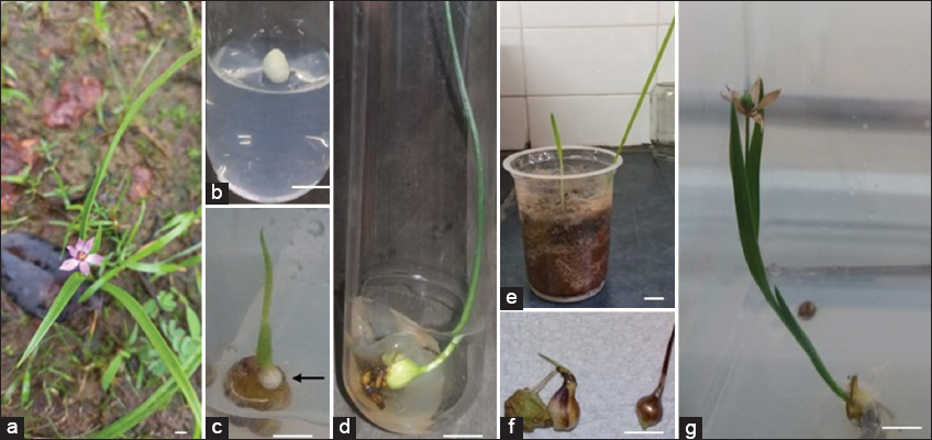 | Figure 2: In vitro propagation of Iphigenia stellata Blatt. using rhizome corm explants: (a) native habitat; (b) shoot initiation with corm bud explant; (c) shoot initiation and induction of microcorms (marked with arrow), (d) in vitro shoot growth and regenerated plants with a well-developed root system; (e) hardening of plantlets; (f) harvesting of microcorms and (g) in vitro flowering in I. stellata. Scale bar = 1 cm. [Click here to view] |
Table 1: Shoot induction response in Iphigenia stellata at different concentrations of BAP, Kn, and TDZ.
| Plant growth regulators (mg/L) | Shoot length (in cm) | Number of shoots per explant with microcorms at the base | ||
|---|---|---|---|---|
| BAP | Kn | TDZ | ||
| 1 | - | - | 7.23±7.25b | 1.33±0.58bc |
| 2 | - | - | 8.13±0.85a | 3.70±0.58a |
| 3 | - | - | 7.3±0.61b | 2.00±1b |
| 4 | - | - | 7.2±0.70b | 1.33±0.58bc |
| 5 | - | - | 6.9±0.1b | 1.33±0.58bc |
| - | 0.1 | - | 0e | f0 |
| - | 0.2 | - | 2.4±4.16d | 0.33±0.58e |
| - | 0.3 | - | 2.5±2.30c | 0.67±0.58d |
| - | 0.4 | - | 6.6±0.51c | 1.00±0c |
| - | 0.5 | - | 5.0±4.36c | 0.67±0.58d |
| - | - | 0.1 | 0e | f0 |
| - | - | 0.2 | 0e | f0 |
| - | - | 0.3 | 0e | f0 |
| - | - | 0.4 | 0e | f0 |
| - | - | 0.5 | 0e | f0 |
BAP: 6-benzylaminopurine, Kn: Kinetin, TDZ: Thidiazuron. Data are presented as mean ± SD. Means sharing a letter in each column do not differ significantly at P < 0.05 (Duncan’s test).
3.2. Root Induction, Hardening, and Acclimatization
Well-developed shoots were transferred onto the root induction medium containing MS fortified with auxins (NAA, IBA, and IAA) at the concentrations mentioned above, of which 0.2 mg/L NAA induced the highest number of roots [Figure 2d]. Nonetheless, maximum root length was observed in the medium with an NAA concentration of 0.5 mg/L [Table 2]. Similar results have been reported for NAA but in combination with IBA in I. indica [18].
Table 2: In vitro root induction response at different concentrations of IBA, IAA and NAA.
| Plant growth regulators (mg/L) | Root length (in cm) | Number of roots per explant | ||
|---|---|---|---|---|
| IBA | IAA | NAA | ||
| 0.1 | - | - | 1.17±1.44de | 0.67±0.58d |
| 0.2 | - | - | 0.73±0.64fghi | 0.67±0.58d |
| 0.3 | - | - | 0.50±0.87fgh | 0.33±0.58ef |
| 0.4 | - | - | 0.57±0.98ghi | 0.33±0.58ef |
| 0.5 | - | - | 1.30±0.1ef | 0.33±0ef |
| - | 0.1 | - | 0j | 0g |
| - | 0.2 | - | 0j | 0g |
| - | 0.3 | - | 0.43±0.75hi | 0.33±0.58ef |
| - | 0.4 | - | 0.90±0.85fg | 0.33±0.58ef |
| - | 0.5 | - | 1.17±1.01ef | 0.33±0.58ef |
| - | - | 0.1 | 2.00±0.58cd | 1.33±0.58bc |
| - | - | 0.2 | 2.03±0.84c | 3.0±1a |
| - | - | 0.3 | 2.17±0.73bc | 2.0±1b |
| - | - | 0.4 | 2.73±0.70b | 1.33±0.58bc |
| - | - | 0.5 | 4.17±1.25a | 1.33±0.58bc |
| - | - | 0.6 | 0.53±0.92 fghi | 0.33±0.58ef |
| - | - | 0.7 | 0.53±0.92ghi | 0.33±0.58ef |
| - | - | 0.8 | 0.43±0.75fghi | 0.33±0.58ef |
| - | - | 0.9 | 0.43±0.75i | 0.33±0.58ef |
| - | - | 1.0 | 0j | 0g |
IBA: Indole-3-butyric acid, IAA: Indole-3-acetic acid, NAA: 1-naphthaleneacetic acid.Data are presented as mean ± SD. Means sharing a letter in each column do not differ significantly at P < 0.05 (Duncan’s test).
IBA and IAA showed non-suitability for corm explants in this study, which is probably due to lower concentrations of auxins and higher alkaloid contents in corm explants, which is likely to interfere with the PGR absorption from the medium, and also possible oxidation of IBA and IAA, reducing their bioavailability. However, further experiments are encouraged to study this phenomenon. Furthermore, a combination of auxins could be investigated for better results.
The regenerated plantlets were replanted in a 1:1:1 mixture of soil, sand, and cocopeat for hardening and transferred to the greenhouse after 1 week [Figure 2e]. All the plantlets survived, showing 100% survival after 1 week. Gradually, the leaves dried out, much like the conditions in the wild. Here, in this study, a new corm arising from a preceding corm suggests clonal multiplication. The corms were collected, shade dried, and preserved in refrigeration at 4°C for further experiments [Figure 2f]. Apparently, the corms showed sprouting in the subsequent experiments, confirming complete viability. The overall shooting and rooting responses of I. stellata corm explants to BAP and NAA, respectively, are consistent with the reports available for I. indica [18] from the Colchicaceae family, indicating the species-specific response.
3.3. In Vitro Flowering Response in the PGR-Free Medium
In this study, in vitro flowering, along with the fruit setting response, was recorded. Several other species have demonstrated a flowering response [20], including Lilium rubellum (Family: Liliaceae), asparagoid lilies Crocus sativus (Family: Iridaceae), and Narcissus bulbocodium (Family: Amaryllidaceae), under in vitro conditions on media supplemented with BAP. However, in I. stellata, the response was observed in PGR-free, half-strength MS medium [Figure 2g] which demonstrates the stress-induced flowering response in PGR-free control medium. No shoot multiplication or microcorm induction was observed in the PGR-free medium, suggesting that it has limitations for in vitro propagation.
The in vitro flowering response in this study was recorded in the winter months, indicating the explant exhibited flowering in a season that is not its ideal flowering season in the natural habitat. Therefore, the flowering response recorded here is indeed a survival response. This in vitro flowering phenomenon can be further studied and utilised to grow, maintain, and propagate I. stellata under in vitro conditions. The continuous year-round maintenance of Iphigenia, along with in vitro flowering and fruit setting under stress-induced conditions, can hence be attributed to the nutrient-deficient media condition, characterized by low nutrient density, and the age of the explant. Few other previously reported in vitro flowering responses highlight a varied range of factors [21] that may induce flowering, including environmental signals and internal factors, which trigger the signal transduction pathways that lead to the conversion of shoot apical meristems into floral meristems. These triggers can be in any form; for example, inherent florigen synthesis, nutrient diversion to the apical meristem, photoperiod variation, and the presence of carbohydrates, nitrogenous compounds, and PGRs can lead to a flowering response [21]. The overall flowering response in I. stellata is exceptional and promising.
In vitro flowering is therefore an important advancement in plant biotechnology, enabling the induction of floral development under controlled laboratory conditions, significantly shortening the breeding cycles, and supporting the propagation of genetically uniform and disease-free plant material [22]. As a result, particularly valuable for the conservation of endangered species, without depleting the natural populations. Furthermore, facilitating the study of detailed physiological and molecular mechanisms of floral development [23], further accelerating plant breeding [24]. Collectively, these attributes underscore the indispensable role of in vitro flowering in fundamental research, plant breeding, and more importantly conservation.
4. CONCLUSION
This research has enabled the development of an efficient protocol for the in vitro propagation of I. stellata Blatt. from corm explants, showing an encouraging response for this endangered species under lab-scale study, also emphasizing the suitability for in vitro flower induction. With further scaling up, this study can be used as basal information for conservation of the endangered species I. stellata as an ex-situ method.
5. ACKNOWLEDGMENTS
The authors are thankful to the KLE Academy of Higher Education and Research, Belagavi, Karnataka, for the funding support received and to the National Institute of Traditional Medicine, ICMR, Belagavi, Karnataka, for providing the laboratory facilities.
6. AUTHORS CONTRIBUTIONS
All authors made substantial contributions to conception and design, acquisition of data, or analysis and interpretation of data; took part in drafting the article or revising it critically for important intellectual content; agreed to submit to the current journal; gave final approval of the version to be published; and agreed to be accountable for all aspects of the work. All the authors are eligible to be an author as per the International Committee of Medical Journal Editors (ICMJE) requirements/guidelines.
7. CONFLICTS OF INTEREST
The authors report no financial or any other conflicts of interest in this work.
8. ETHICAL APPROVALS
This study does not involve experiments on animals or human subjects.
9. DATA AVAILABILITY
All the data is available with the authors and shall be provided upon request.
10. PUBLISHER’S NOTE
All claims expressed in this article are solely those of the authors and do not necessarily represent those of the publisher, the editors and the reviewers. This journal remains neutral with regard to jurisdictional claims in published institutional affiliation.
11. USE OF ARTIFICIAL INTELLIGENCE (AI)-ASSISTED TECHNOLOGY
The authors declare that they have not used artificial intelligence (AI)-tools for writing and editing of the manuscript, and no images were manipulated using AI..
REFERENCES
1. Yadav SR, Sardesai MM. Flora of Kolhapur District. Maharashtra:Shivaji University Kolhapur; 2002:499-500.
2. Patel MB. Star grass lily Iphigenia stellata blatter (Colchicaceae)-a new addition to the flora of Gujarat, India. J Threatened Taxa. 2021;13(1):17604-6.[CrossRef]
3. Sharma BD, Karthikeyan S, Singh NP, Lakshminarasimhan P. Flora of India Series 2, Flora of Maharashtra State Monocotyledons. Kolkata: Botanical Survey of India;1996. 140.
4. Lekhak MM, Surveswaran S, Yadav SR. Generic identity of Camptorrhiza indica (Colchicaceae) based on cytogenetics and molecular phylogenetics. J Syst Evol. 2016;54(1):75-82.[CrossRef]
5. World Flora Online. Iphigenia. WFO Plant List;2022. Available from: https://wfoplantlist.org/taxon/wfo-4000019149-2022-12?page=1 [Last accessed on 2025 Apr 20].
6. IUCN. Iphigenia ratnagirica. The IUCN Red List of Threatened Species;2024. Available from: https://www.iucnredlist.org/species/191675230/191678437 [Last accessed on 2025 Apr 20].
7. Sadiq NM, Robinson KJ, Terrell JM. Colchicine. In:StatPearls. Treasure Island, FL:StatPearls Publishing;2025. Available from: https://www.ncbi.nlm.nih.gov/books/NBK431102 [Last accessed on 2025 Apr 20].
8. Ade R, Rai MK. Review:Colchicine, current advances and future prospects. Nusantara Biosci. 2010;2(2):90-6.[CrossRef]
9. Colchicine:Uses, Dosage, Side Effects, Warnings. Available from: https://www.drugs.com/colchicine.html [Last accessed on 2025 Apr 20].
10. IUCN. Iphigenia stellata. IUCN Red List of Threatened Species;2015. Available from: https://www.iucnredlist.org/details/50126619/0 [Last accessed on 2025 Apr 20].
11. Plant Cell Technology. Cytokinin and Shoot Development. Plant Cell Technology Blog;2024. Available from: https://plantcelltechnology.com/blogs/blog/blog-cytokinin-and-shoot-development [Last accessed on 2025 Apr 20].
12. Plant Cell Technology. Auxin:The Rooting Hormone. Plant Cell Technology Blog;2024. Available from: https://plantcelltechnology.com/blogs/blog/blog-auxin-the-rooting-hormone [Last accessed on 2025 Apr 20].
13. Murashige T, Skoog F. A revised medium for rapid growth and bio assays with tobacco tissue cultures. Physiol Plant. 1962;15(3):473-97.[CrossRef]
14. Kulak V, Longboat S, Brunet ND, Shukla M, Saxena P. In vitro technology in plant conservation:Relevance to biocultural diversity. Plants. 2022;11(4):503.[CrossRef]
15. Thakur J, Abbas NS, Bhardwaj S, Kaula BC. Biotechnological tools in the propagation and conservation of threatened species:An overview. IJBT. 2021;20:217-35.
16. Komakech R, Kim YG, Kim WJ, Omujal F, Yang S, Moon BC, et al. A micropropagation protocol for the endangered medicinal tree Prunus africana (Hook f.) Kalkman:Genetic fidelity and physiological parameter assessment. Front Plant Sci. 2020;11:548003.[CrossRef]
17. Zabicka J, Zabicki P, Slomka A, Sliwinska E, Jedrzejczyk-Korycinska M, Nowak T, et al. Re-introduction of an extinct population of Pulsatilla patens using different propagation techniques. Sci Rep. 2022;12(1):14321. [CrossRef]
18. Mukhopadhyay MJ, Mukhopadhyay S, Sen S. In vitro propagation of Iphigenia indica, an alternative source of colchicine. Plant Cell Tissue Organ Cult. 2002;69:101-4.[CrossRef]
19. Mukhopadhyay MJ, Mukhopadhyay S. High frequency in vitro propagation through somatic embryogenesis of Iphigenia indica Kunth et Benth, an endangered medicinal and colchicines yielding herb of commercial interest. Cytologia. 2008;73(2):97-103.[CrossRef]
20. Murthy KS, Kondamudi R, Rao PC, Pullaiah T. In vitro flowering-a review. J Agric Technol. 2012;8(5):1517-36.
21. Sudhakaran S, Teixeira da Silva JA, Sreeramanan S. Test Tube Bouquets:In vitro Flowering. Floriculture, Ornamental and Plant Biotechnology: Advances and Topical Issues. Vol. 2. Isleworth:Global Science Books;2006. 336-46.
22. Kaur S. In vitro florigenesis with special reference to orchids-a review. Recent Pat Biotechnol. 2022;16(4):311-8.[CrossRef]
23. Sharma V, Kamal B, Srivastava N, Dobriyal AK, Jadon VS. In vitro flower induction from shoots regenerated from cultured axillary buds of endangered medicinal herb Swertia chirayita H. Karst. Biotechnol Res Int. 2014;2014(1):264690.[CrossRef]
24. Shen P, Gao S, Hu J, Li Y, Lei T, Shi L. In vitro flowering of the distylous plant Plumbago auriculata Lam. South Afr J Bot. 2021;137:492-8.[CrossRef]