
1. INTRODUCTION
Antimicrobial resistance (AMR) is recognized as a major global public health concern of the twenty-first century [1,2]. Although clinical settings have historically been considered the primary source of multidrug-resistant (MDR) bacteria, increasing evidence indicates that AMR is widely distributed across environmental and food-associated ecosystems [3,6]. Aquatic environments, in particular, are increasingly recognized as important reservoirs and transmission pathways for AMR due to continuous exposure to anthropogenic activities, including the use of antibiotics in agriculture and aquaculture, discharge of untreated effluents, and natural microbial interactions [4-8]. These factors collectively contribute to the persistence, evolution, and dissemination of resistant bacteria beyond clinical boundaries [3,5,7].
The gastrointestinal tract of fish represents a complex and dynamic microbial ecosystem in which resident and transient microorganisms interact under various selective pressures, such as diet, environmental conditions, and microbial competition [9]. Even in the absence of visible disease, fish may harbor bacteria carrying AMR and virulence determinants, thereby acting as asymptomatic reservoirs [10]. Fish intended for human consumption are of particular concern, as intestinal bacteria may enter the food chain during handling, processing, or preparation [11]. Therefore, the investigation of AMR bacteria in apparently healthy fish obtained from retail markets is essential for understanding potential pathways of resistance transmission at the human–animal–environment interface.
Catla catla, commonly known as catla, is one of the major Indian carps widely cultured in South Asia due to its rapid growth rate, high nutritional value, and economic importance in aquaculture [12]. It is an important component of freshwater aquaculture systems in countries such as India, Bangladesh, and Pakistan and contributes substantially to regional fish production [13]. Owing to its widespread cultivation and high market demand, C. catla is frequently exposed to intensive aquaculture practices, including the use of antibiotics for disease prevention and therapeutic purposes [4,14]. Such practices can promote the selection and persistence of AMR bacteria within the fish microbiota [14]. Moreover, fish markets represent critical points of interaction between aquatic organisms, environmental contaminants, and human populations, facilitating the potential transfer of microbial communities [15]. The intestinal microbiota of C. catla may harbor diverse bacterial populations, including opportunistic and AMR species, which can enter the food chain during handling and processing [16]. Therefore, studying bacterial isolates from C. catla is important for understanding the role of freshwater fish in the dissemination of AMR within the One Health framework.
Staphylococcus haemolyticus is a Gram-positive, coagulase-negative bacterium that forms part of the normal microbiota of humans and animals. Although traditionally considered a commensal organism, it has emerged as an opportunistic pathogen with increasing clinical relevance due to its ability to acquire resistance to multiple antimicrobial agents. Staphylococcus haemolyticus is now recognized as an important cause of nosocomial infections, including bacteremia, septicemia, and device-associated infections, particularly in immunocompromised individuals [17]. This species has been reported from a wide range of sources, including clinical environments, animals, food products, and aquatic ecosystems, highlighting its zoonotic potential and significance within the One Health framework [18]. Its widespread distribution, high genomic plasticity, and capacity to harbor AMR determinants suggest that it may act as an important reservoir of resistance genes and facilitate their transfer to other bacterial species. In addition, pathogenic strains of S. haemolyticus are characterized by their ability to form biofilms and produce virulence-associated factors, which enhance survival, persistence, and resistance in both clinical and environmental settings [17].
Whole-genome sequencing (WGS) has emerged as a powerful tool for investigating the genetic basis of AMR and understanding bacterial adaptation. In the present study, a MDR S. haemolyticus isolate was recovered from the intestinal tract of C. catla obtained from a local fish market. To our knowledge, this represents one of the few reports describing WGS-based characterization of MDR S. haemolyticus from freshwater fish in India. The isolate was characterized using phenotypic antimicrobial susceptibility testing, 16S ribosomal RNAs (rRNA) gene sequencing, and WGS to investigate its genomic features, AMR genes, and virulence determinants. This study aims to enhance the understanding of AMR in aquatic food systems and highlights the importance of monitoring food-associated bacteria within a One Health framework.
2. MATERIALS AND METHODS
2.1. Sample Collection and Isolation
Samples of C. catla were collected from JCT Fish Market, Phagwara, Jalandhar, Punjab, India, during October 2025. Fish were not selected based on visible clinical signs of disease and were sampled opportunistically as part of an exploratory assessment of AMR bacteria associated with market-sold fish. At the time of sampling, the fish were dead and, according to vendors, had been displayed at the market for several hours prior to collection. Samples were transported to the laboratory under ice-cold conditions. Intestinal contents were aseptically removed, homogenized, serially diluted up to 10-5 in sterile saline, and 100 μl from each dilution was spread onto nutrient agar (NA) plates and incubated aerobically. Distinct colonies were subcultured for purification. The inoculated plates were incubated at 29°C ± 1°C for 24 hours. Following incubation, morphologically distinct colonies were carefully selected and purified through successive streaking on fresh NA plates to obtain pure (axenic) cultures. Colony differentiation was performed based on morphological characteristics such as size, shape, margin, elevation, and pigmentation. Each purified isolate was subjected to Gram staining and a series of standard biochemical assays, including catalase, oxidase, coagulase, indole production, urease activity, nitrate reduction, motility, mannitol fermentation, oxidation/fermentation, and hemolysis on blood agar, following conventional microbiological procedures [19,20]. Pure cultures were preserved in nutrient broth supplemented with 20% (v/v) glycerol and stored at −80°C. Based on phenotypic identification, a total of 20 distinct bacterial isolates were obtained from five fish samples and subsequently evaluated for antimicrobial susceptibility.
2.2. Antibiotic Susceptibility Testing (AST)
Antimicrobial susceptibility was determined using the Kirby–Bauer disk diffusion method on Mueller–Hinton agar (MHA) in accordance with the Clinical and Laboratory Standards Institute (CLSI, 2019) guidelines [21]. Briefly, bacterial suspensions were prepared from overnight cultures and adjusted to a 0.5 McFarland turbidity standard. The standardized inoculum was uniformly spread onto MHA plates using sterile swabs, and antibiotic discs were aseptically placed on the agar surface. The antibiotics evaluated included imipenem (10 µg), tetracycline (30 µg), ofloxacin (5 µg), ciprofloxacin (5 µg), gentamicin (10 µg), levofloxacin (5 µg), norfloxacin (10 µg), amoxicillin (10 µg), erythromycin (15 µg), tobramycin (10 µg), cefotaxime (10 µg), and chloramphenicol (30 µg). These antibiotics were selected to represent diverse antimicrobial classes, including β-lactams, aminoglycosides, fluoroquinolones, tetracyclines, macrolides, phenicols, and carbapenems, based on their clinical importance in human and veterinary medicine and their relevance in AMR surveillance studies. All antibiotic discs were procured from HiMedia Laboratories Pvt. Ltd., Mumbai, India.
Plates were incubated at 35°C for 16–18 hours. Zones of inhibition were measured in millimeters and interpreted as susceptible, intermediate, or resistant according to CLSI M100 Performance Standards for Antimicrobial Susceptibility Testing (2019) and the corresponding breakpoint criteria for staphylococci. For β-lactam antibiotics (amoxicillin, cefotaxime, and imipenem), susceptibility interpretations were based on archived CLSI M100 breakpoint criteria previously established for Staphylococcus spp [22]. The test was conducted in triplicate to ensure reproducibility. Isolates exhibiting resistance to three or more classes of antibiotics were classified as MDR [23]. The isolate demonstrating resistance to the highest number of antimicrobial agents was selected for subsequent molecular identification and WGS analysis.
2.3. 16S rRNA Gene Sequencing
Genomic DNA was extracted from the bacterial isolate using the Qiagen DNeasy UltraClean Microbial Kit in accordance with the manufacturer's protocol. The integrity of the extracted DNA was verified by electrophoresis on a 0.8% agarose gel at 110 V for 30 minutes. DNA purity and concentration were determined spectrophotometrically using a BioTek Epoch microplate reader by measuring the absorbance ratio at 260/280 nm.
The 16S rRNA gene was amplified by polymerase chain reaction (PCR) using universal primers 27F and 1391R [24]. PCR products were visualized by agarose gel electrophoresis and subsequently purified using a column-based purification kit to remove contaminants. Sequencing of the purified amplicons was performed using the BigDye Terminator v3.1 Cycle Sequencing Kit on an ABI 3500xl Genetic Analyzer. The obtained sequences were compared against reference sequences in the NCBI GenBank database using the BLAST algorithm to determine species-level identification.
2.4. WGS and Assembly
Sequencing libraries were prepared using the TWIST Library Preparation Kit 2.0 (Twist Bioscience, USA) following the manufacturer's protocol. The procedure included DNA fragmentation, end-repair, adapter ligation, purification, amplification, and quantification steps. Library quality and fragment size distribution were verified using the Agilent 2100 Bioanalyzer (Agilent Technologies, USA).
WGS was performed on the MGI DNBSEQ-G400 platform (MGI Tech Co., Ltd., China) using paired-end chemistry (2 × 150 bp).
2.5. Quality Control and Genome Assembly
Raw paired-end reads were subjected to adapter trimming and quality filtering using Cutadapt and Trim Galore, with a Phred quality score threshold of Q20. High-quality reads were de novo assembled using SPAdes v3.13.0 [25]. Contigs shorter than 500 bp were excluded from downstream analyses. Assembly statistics were generated using QUAST [26]. Genome completeness and contamination were assessed using CheckM v1.2.1 [27].
2.6. Genome Annotation and Functional Characterization
The assembled genome was annotated using the Bacterial and Viral Bioinformatics Resource Center (BV-BRC) (PATRIC) RASTtk [28] pipeline and further validated using Bakta version 1.9.2 [29]. Coding sequences (CDSs), transfer RNAs (tRNAs), rRNAs, and other genomic features were identified during the annotation process.
Functional annotation was performed by mapping predicted protein sequences against the Kyoto Encyclopedia of Genes and Genomes (KEGG) [30] database using BlastKOALA. KEGG Mapper was subsequently used to reconstruct metabolic pathways.
Gene Ontology assignments were obtained using the PANTHER [31] Classification System, and Clusters of Orthologous Groups (COG) classification was performed using COGclassifier [32]. Circular genome visualization was generated using GenoVi version 0.4.3 [33].
2.7. Phylogenetic Analysis
Phylogenetic analysis was performed to determine the evolutionary relationship of the isolate with closely related bacterial strains using a whole-genome-based approach. The phylogenetic tree was constructed using the Codon Tree method implemented in the BV-BRC platform [28].
Initially, closely related reference genomes were selected using the Similar Genome Finder service available within the BV-BRC server. A set of approximately 40–50 closely related genomes was chosen based on genomic similarity to the isolate. The Codon Tree pipeline identifies single-copy orthologous genes (PGFams) conserved across the selected genomes. These genes were extracted and aligned at both the protein and nucleotide levels. The aligned sequences were concatenated to generate a comprehensive dataset representing conserved genomic regions.
Phylogenetic relationships were inferred using the maximum-likelihood (ML) method implemented in Randomized Axelerated Maximum Likelihood (RAxML) [34]. The analysis was performed using appropriate substitution models, and branch support values were estimated using bootstrap analysis to ensure reliability of the inferred phylogeny. The resulting phylogenetic tree was visualized and exported in Scalable Vector Graphics format and further processed using tree visualization tools such as FigTree for presentation. The final tree illustrates the clustering pattern of the isolate with reference strains, enabling accurate determination of its evolutionary position.
2.8. Identification of AMR, Virulence, and Plasmid Genes
AMR genes were identified by screening the assembled genome against the ResFinder (v4.2) and CARD (Resistance Gene Identifier) databases [35,36]. In addition, genome annotation performed using the BV-BRC platform was used to identify and classify resistance-associated genes based on the PATRIC, CARD, and NDARO resistance databases. Virulence-associated genes were detected using Abricate against the Virulence Factor Database (VFDB) and the BV-BRC virulence factor pipeline [28,37]. Putative plasmid sequences were identified using PlasmidFinder (v2.1) with minimum identity and coverage thresholds of 95% and 60%, respectively [38]. Additional plasmid confirmation was performed using the PLSDB database [39].
2.9. Prediction of Pathogenic Potential
The pathogenic potential of the isolate was assessed using PathogenFinder to predict its likelihood of being a human-associated pathogen based on genomic signatures [40].
2.10. Comparative Genomic Analysis Average Nucleotide Identity (ANI)
Comparative genomic analysis was performed using ANI to evaluate the genomic similarity of isolate CGPA with closely related reference genomes [41]. The assembled genome sequence of the isolate was compared against publicly available reference genomes retrieved from the NCBI database.
In addition, the Similar Genome Finder service of the BV-BRC (PATRIC) platform was utilized to identify closely related genomes based on genomic distance estimation using Mash (v2.3) [28,42]. The results obtained from the ANI analysis were further verified through manual comparison with selected reference genomes to ensure accuracy and consistency of genomic similarity estimates.
3. RESULTS
3.1. Antimicrobial Susceptibility Profile and Biochemical Characterization
Among the total bacterial isolates recovered, isolate CGPA exhibited a MDR phenotype. Antimicrobial susceptibility testing was performed using the Kirby–Bauer disk diffusion method, which revealed resistance to eight antibiotics representing different antimicrobial classes. The isolate exhibited complete resistance, characterized by the absence of an inhibition zone (0 mm), against amoxicillin (10 µg), chloramphenicol (30 µg), ciprofloxacin (5 µg), norfloxacin (10 µg), ofloxacin (5 µg), and tetracycline (30 µg). A markedly reduced inhibition zone (2 mm) against erythromycin (15 µg). Additionally, the isolate exhibited a reduced zone of inhibition against imipenem (10 µg). Interpretations of β-lactam antibiotics (amoxicillin, cefotaxime, and imipenem) were based on archived CLSI M100 breakpoint criteria for Staphylococcus spp. These findings confirm the MDR nature of isolate CGPA and highlight its resistance to several clinically important antimicrobial agents. In contrast, the isolate remained susceptible to cefotaxime (10 µg), gentamicin (10 µg), tobramycin (10 µg), and levofloxacin (5 µg), demonstrating measurable zones of inhibition consistent with susceptibility standards. The observed resistance pattern indicates that CGPA possesses resistance against β-lactams, fluoroquinolones, tetracyclines, and phenicols, thereby fulfilling the criteria for MDR. Based on its resistance profile, CGPA was selected for further molecular characterization and WGS analysis. The detailed disk diffusion antimicrobial susceptibility results, including inhibition zone diameters and interpretive categories, are summarized in Table 1.
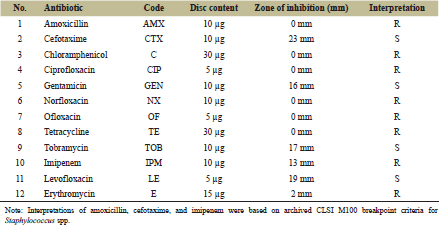 | Table 1. Antimicrobial susceptibility profile of S. haemolyticus isolate CGPA determined by the Kirby–Bauer disk diffusion method. [Click here to view] |
The biochemical characteristics of isolate CGPA were consistent with those reported for S. haemolyticus and were used as preliminary phenotypic indicators prior to molecular confirmation. The isolate exhibited Gram-positive cocci morphology and demonstrated biochemical reactions typical of the genus Staphylococcus. The results of Gram staining and biochemical tests are summarized in Table 2. Biochemical characterization was performed solely for initial phenotypic assessment and was not used as the sole criterion for species-level identification. Definitive identification was subsequently confirmed through 16S rRNA gene sequencing and whole-genome-based analyses.
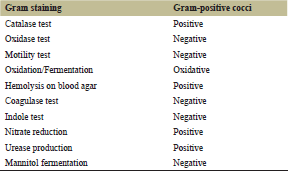 | Table 2. Morphological and biochemical characteristics of S. haemolyticus CGPA. [Click here to view] |
3.2. Molecular Identification (16S rRNA Analysis)
The 16S rRNA gene sequence analysis of isolate CGPA using BLASTn revealed 100% query coverage and up to 99.69% sequence identity with S. haemolyticus. The top BLAST hits included S. haemolyticus strain JCM 2416 and other closely related Staphylococcus species, all showing high sequence similarity. The extremely low E-value (approaching zero) indicated a highly significant match, confirming the reliability of the sequence alignment. Based on nucleotide homology and phylogenetic analysis, the isolate was identified as S. haemolyticus.
3.3. Genome Assembly and Genomic Features
WGS of isolate CGPA generated a total of 26,671,642 paired-end reads, producing high-quality sequencing data with Q30 values exceeding 97%, indicating excellent base-calling accuracy. After quality filtering and trimming, the processed reads were used for de novo assembly.
The draft genome assembly of S. haemolyticus CGPA resulted in a total genome size of 2,565,197 bp (2.57 Mb) with a Guanine–Cytosine (GC) content of 32.71%, consistent with previously reported genomes of S. haemolyticus. The assembly comprised 177 contigs (≥500 bp), with the largest contig measuring 142,991 bp. The contig N50 value was 38,787 bp, and the L50 value was 18, indicating moderate assembly contiguity typical of short-read draft genomes. No ambiguous bases (Ns) were detected in the final assembly.
Genome quality assessment using CheckM demonstrated 100% completeness, with coarse and fine consistency values of 99.9% and 99.3%, respectively, confirming the high reliability and integrity of the assembled genome.
3.4. Genome Annotation
Genome annotation using the BV-BRC (RASTtk) platform predicted a total of 2,667 genes, including 2,608 protein-CDSs, 53 tRNA genes, and 6 rRNA genes. Among the predicted proteins, 2,142 proteins were assigned functional annotations, whereas 466 proteins were classified as hypothetical proteins.
Functional classification revealed that a large proportion of genes were involved in metabolic pathways, cellular processes, and environmental response mechanisms. Subsystem analysis indicated that most genes were associated with metabolic functions, protein processing, stress response, and cellular respiration. The distribution of genes across functional subsystems is presented in Figure 1. The detailed assembly characteristics, genomic features, and taxonomic classification of the isolate are summarized in Table 3.
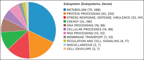 | Figure 1. Subsystem distribution of genes in S. haemolyticus CGPA genome based on BV-BRC (RASTtk) functional annotation. [Click here to view] |
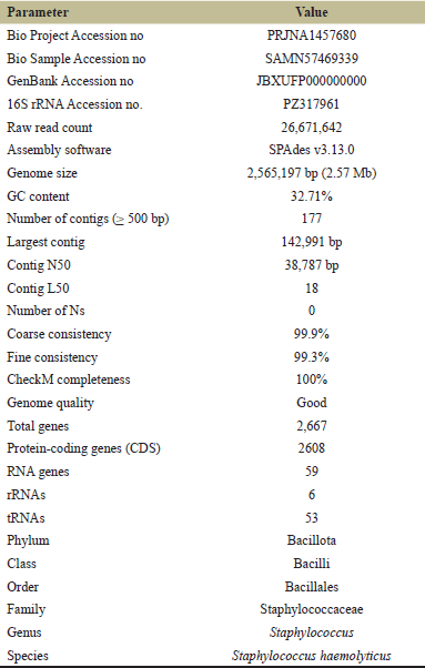 | Table 3. Assembly characteristics and taxonomy of the annotated genome. [Click here to view] |
3.5. Functional Annotation and Subsystem Analysis
Functional annotation of the assembled genome of S. haemolyticus isolate CGPA was performed using the BV-BRC (RASTtk) pipeline and further validated with Bakta. A total of 2,667 genes were predicted in the genome, including 2,608 protein-CDSs and 59 RNA genes. Among the predicted proteins, 2,142 genes were assigned putative functional roles, whereas 466 genes were annotated as hypothetical proteins, indicating the presence of several proteins with currently unknown biological functions.
Subsystem analysis revealed that the majority of annotated genes were associated with metabolic and cellular processes, reflecting the organism's metabolic versatility and adaptability to diverse environmental conditions. The identified subsystems were primarily involved in carbohydrate metabolism, amino acid metabolism, energy production, and cofactor and vitamin metabolism.
3.6. KEGG Pathway Annotation
Functional annotation of the genome using the KEGG database identified 920 KEGG orthologs in the genome of S. haemolyticus CGPA. These orthologs were distributed across multiple metabolic and cellular pathways, highlighting the metabolic versatility of the organism.
Among the annotated pathways, carbohydrate metabolism constituted the largest functional category, comprising 206 genes, followed by amino acid metabolism (152 genes), metabolism of cofactors and vitamins (135 genes), and energy metabolism (102 genes). Additional pathways included nucleotide metabolism, lipid metabolism, and glycan biosynthesis, indicating the presence of diverse biochemical processes required for cellular maintenance and adaptation. The KEGG-based pathway reconstruction demonstrates the presence of complex metabolic networks supporting bacterial survival and environmental adaptability. The distribution of KEGG pathway categories is illustrated in Figure 2.
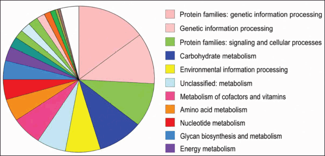 | Figure 2. Functional categorization of genes in S. haemolyticus CGPA based on KEGG pathway annotation. [Click here to view] |
3.7. COG Functional Classification
COG classification was performed to further categorize predicted proteins based on functional roles. The COG analysis revealed that a large proportion of genes were associated with metabolic functions, cellular processes, and information storage and processing. The most prominent categories included amino acid transport and metabolism, carbohydrate transport and metabolism, transcription, and translation-related functions, indicating active genetic regulation and metabolic activity within the genome.
Additional functional groups included energy production and conversion, cell wall and membrane biogenesis, and signal transduction mechanisms, reflecting the physiological complexity of the organism. Several genes were also assigned to the "general function prediction only" and "unknown function" categories, suggesting the presence of proteins with yet uncharacterized biological roles. Overall, the COG classification highlights the diverse functional repertoire of the CGPA genome and its capacity to adapt to different environmental conditions. The overall distribution of genes across COG functional categories is presented in Figure 3.
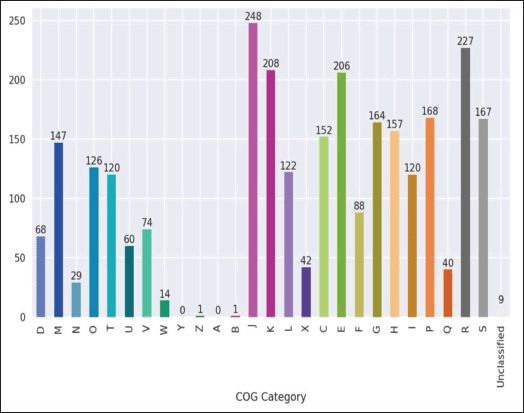 | Figure 3. Functional categorization of genes in S. haemolyticus CGPA based on COG classification. [Click here to view] |
3.8. Circular Genome Representation
A circular genome map of the draft genome of S. haemolyticus isolate CGPA was generated using the GenoVi visualization tool to illustrate the structural organization of genomic features. The circular representation displays multiple genomic layers, including the distribution of protein-coding genes, RNA genes, GC content, and GC skew across the assembled genome. CDSs located on the forward and reverse strands are represented in separate rings, highlighting gene orientation and distribution along the genome.
Additional layers of the genome map depict variations in GC content and GC skew, which provide insights into genomic composition and replication dynamics. The visualization also highlights the overall organization of genomic regions within the assembled contigs. The circular genome representation provides an overview of the structural architecture of the CGPA genome and facilitates the visualization of gene distribution and genomic features. The complete circular genome map is presented in Figure 4.
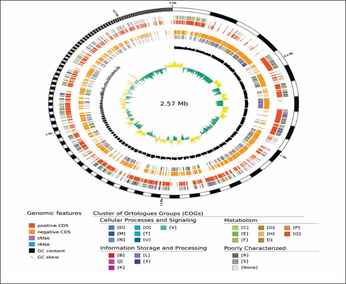 | Figure 4. Circular genome representation of S. haemolyticus CGPA generated using the GenoVi visualization tool. Rings represent CDSs on the forward and reverse strands, GC content variation, and GC skew across the genome. [Click here to view] |
3.9. Phylogenetic Analysis
Phylogenetic analysis was performed using the Codon Tree pipeline implemented in the BV-BRC server to determine the evolutionary placement of the CGPA isolate among related members of the genus Staphylococcus. Reference and representative genomes were automatically selected by the BV-BRC database for comparative analysis. Phylogenetic reconstruction was performed using conserved protein families (PGFams). Protein sequences were aligned using MUSCLE, and the corresponding nucleotide sequences were mapped onto the protein alignments. The combined amino acid and nucleotide alignments were concatenated into a single data matrix, which was analyzed using the ML algorithm implemented in RAxML.
Bootstrap analysis was performed to assess the statistical support for each branch (Fig. 5). The resulting phylogenetic tree demonstrated that isolate CGPA clustered closely with S. haemolyticus strain JCSC1435, forming a well-supported clade with a bootstrap value of 100. This clustering clearly separated the CGPA isolate from other closely related Staphylococcus species, including Staphylococcus epidermidis, S. aureus, and Staphylococcus warneri. These findings further confirm the taxonomic identification of the isolate as S. haemolyticus and indicate a strong evolutionary relationship with previously characterized strains.
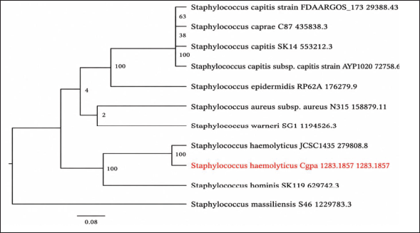 | Figure 5. ML phylogenetic tree showing the relationship of isolate CGPA with representative Staphylococcus species, constructed using the Codon Tree pipeline in BV-BRC based on PGFams. Bootstrap values are indicated at the nodes. Isolate CGPA (highlighted in red) clustered with S. haemolyticus strain JCSC1435, confirming its species identity. [Click here to view] |
3.10. Antibiotic Resistance Genes
AMR gene analysis was performed using ResFinder, CARD (RGI), and the BV-BRC annotation platform. ResFinder did not identify any acquired AMR genes. CARD analysis detected several high-confidence resistance determinants, including mecA, APH(3′)-IIIa, ANT(4′)-Ia, qacA, blaI, blaR1, erm(C), tet(K), tet(L), vgaALC, and catA8, which are associated with resistance to β-lactams, aminoglycosides, macrolides, tetracyclines, phenicols, and antiseptic compounds. The differences observed between ResFinder and CARD results likely reflect differences in database composition, curation criteria, and the types of resistance determinants targeted by each platform. ResFinder primarily identifies acquired AMR genes, whereas CARD and BV-BRC additionally detect resistance-associated determinants and chromosomal resistance mechanisms. In addition, BV-BRC annotation identified resistance-associated genes across the PATRIC, CARD, and NDARO databases. Collectively, CARD and BV-BRC identified APH(3′)-IIIa, vgaALC, blaI/blaR1, qacA, erm(C), ANT(4′)-Ia, tet(45), tet(K), tet(L), mecA, catA8, and PC1 as resistance-associated determinants. The identified resistance genes and their associated resistance mechanisms are summarized in Table 4. In addition to the resistance genes identified by CARD and BV-BRC, functional classification of AMR-related genes and antibiotic target genes identified through BV-BRC annotation is presented in Table 5. These genes include antibiotic targets, efflux-associated proteins, regulatory elements, and resistance-associated functional categories that may contribute to antimicrobial adaptation.
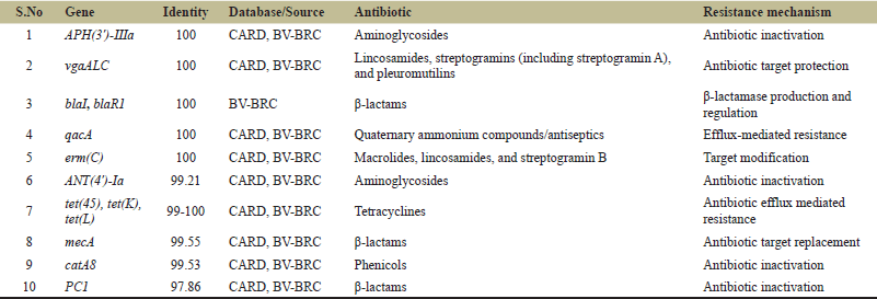 | Table 4. AMR genes identified in S. haemolyticus isolate CGPA using CARD and BV-BRC databases. [Click here to view] |
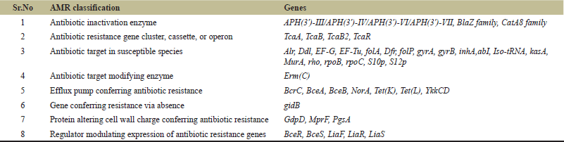 | Table 5. AMR-related genes and antibiotic target genes identified through BV-BRC annotation. [Click here to view] |
3.11. Virulence Factors
Virulence factor analysis identified the presence of the clpP gene, encoding an ATP-dependent Clp protease associated with stress survival and pathogenicity. This gene showed high query coverage (97%) and sequence identity (80%), suggesting its functional significance in the isolate.
3.12. Plasmid Analysis
Plasmid analysis using PlasmidFinder did not resolve any complete plasmid sequence within the draft genome assembly. However, plasmid replicon-associated sequences, including rep5b, rep7a, rep10, and rep20, were detected. These replicon sequences are summarized in Table 6.
 | Table 6. Plasmid replicon sequences identified in S. haemolyticus CGPA using PlasmidFinder. [Click here to view] |
To further validate the plasmid-associated sequences, the assembled genome was screened against the PLSDB database. The analysis identified 127 significant matches to previously reported circular plasmids, including plasmids associated with Staphylococcus species, with sequence identities ranging from 99.2% to 99.98%. These findings further support the presence of plasmid-associated elements within the genome assembly.
3.13. PathogenFinder Analysis
PathogenFinder analysis predicted the isolate to be a human pathogen with a probability score of 0.97, indicating high pathogenic potential.
3.14. Comparative Genomic Analysis (ANI)
Comparative genomic analysis using ANI revealed a high level of genomic similarity between isolate CGPA and reference strains of S. haemolyticus. ANI values ranged from 99.27% to 99.31%, with the highest similarity observed with strain ASM799279v1 (99.311%), followed by ASM609439v1 (99.281%), ASM290180v1 (99.274%), ASM2902394v1 (99.277%), and strain 36734_F01 (99.276%). These values are significantly above the accepted species delineation threshold of 95%, confirming that isolate CGPA belongs to S. haemolyticus. The consistently high ANI values indicate strong genomic conservation and close evolutionary relatedness among the analyzed strains.
4. DISCUSSION
AMR is increasingly recognized across environmental and food-associated ecosystems, with aquatic systems acting as important reservoirs and transmission interfaces within a One Health framework. Continuous exposure to antibiotics from aquaculture practices, agricultural runoff, and wastewater inputs promotes the selection and persistence of resistant bacteria in these environments [4,6]. In this context, the isolation of a MDR S. haemolyticus from the intestinal tract of C. catla obtained from a retail fish market highlights a potential route for the dissemination of AMR through the fish food chain.
Similar findings have been reported in aquaculture systems, where S. haemolyticus has been identified as a potential emerging pathogen in fish species. A previous study reported its involvement in disease outbreaks in farmed fish, where infected fish exhibited clinical signs such as anorexia, abnormal swimming behavior, ulceration, and internal organ congestion, with experimental infection confirming its pathogenic potential [43]. These observations support the present findings and suggest that S. haemolyticus may act as an opportunistic pathogen in aquatic environments.
Phenotypically, isolate CGPA exhibited resistance to multiple antibiotic classes, including β-lactam agents evaluated in the present study, fluoroquinolones, tetracyclines, and phenicols. Such broad resistance profiles are consistent with previous reports identifying S. haemolyticus as one of the most resistant coagulase-negative staphylococci, with a notable capacity to accumulate resistance determinants [44,45]. The MDR observed in isolate CGPA is in agreement with previous studies reporting methicillin-resistant S. haemolyticus in retail fish samples [46].
However, methicillin resistance was not phenotypically confirmed in the present isolate. In addition, interpretations of β-lactam antibiotics (amoxicillin, cefotaxime, and imipenem) were based on archived CLSI M100 breakpoint criteria for Staphylococcus spp. These findings suggest that fish and fish-associated environments can act as reservoirs of MDR bacteria. The occurrence of MDR S. haemolyticus in a food-associated source underscores the ecological versatility of this species and supports its role as a reservoir of resistance genes beyond clinical settings.
Molecular identification based on 16S rRNA gene sequencing showed high sequence similarity (≥99%) with S. haemolyticus, consistent with established approaches for bacterial identification [24]. Whole-genome-based phylogenetic analysis further confirmed this classification, and ANI values exceeding 99% supported robust species delineation [47].
WGS provided detailed insight into the genetic basis of the MDR phenotype. Notably, the detection of the mecA gene indicates the genetic potential for methicillin resistance through the production of PBP2a, a low-affinity penicillin-binding protein [48]. However, phenotypic confirmation using cefoxitin or oxacillin susceptibility testing was not performed in the present study. In staphylococci, mecA is typically associated with the staphylococcal cassette chromosome mec (SCCmec), a mobile genetic element responsible for the horizontal transfer of methicillin resistance. However, SCCmec elements were not explicitly identified in the present study, suggesting that further genomic investigation is required to confirm their genomic context. The detection of mecA in a food-associated isolate highlights the potential for environmental dissemination of clinically relevant resistance determinants.
Although the aminoglycoside resistance genes APH(3′)-IIIa and ANT(4′)-Ia were detected in isolate CGPA, the isolate remained phenotypically susceptible to gentamicin and tobramycin. APH(3′)-IIIa and ANT(4′)-Ia encode aminoglycoside-modifying enzymes that have been associated with aminoglycoside resistance mechanisms in bacteria [49]. However, the presence of these genes was not reflected in the phenotypic susceptibility profile observed in the present study. Resistance to tetracycline was associated with the presence of tet(45), tet(K), and tet(L), which are known to mediate efflux-based resistance mechanisms [50]. Furthermore, the detection of qacA, an efflux pump-associated gene involved in resistance to quaternary ammonium compounds and antiseptics, highlights the presence of additional efflux-mediated resistance mechanisms within the genome [51]. Additionally, the presence of catA8 explains resistance to phenicol antibiotics through enzymatic inactivation [52]. Although phenotypic resistance to ciprofloxacin, norfloxacin, and ofloxacin was observed, specific fluoroquinolone resistance-associated mutations in gyrA, parC, grlA, or grlB were not investigated in the present study. Therefore, the genetic basis of fluoroquinolone resistance in isolate CGPA could not be conclusively determined and warrants further investigation.
The coexistence of multiple resistance mechanisms, including enzymatic inactivation, target modification, and active efflux, highlights the complex and multifactorial nature of AMR in this isolate.
From a virulence perspective, the identification of the clpP gene provides insight into the adaptive and pathogenic potential of the isolate. ClpP is involved in protein quality control, stress tolerance, and regulation of cellular processes in Gram-positive bacteria [53,54]. Previous studies have shown that the Clp protease system contributes to stress adaptation and virulence regulation in several Gram-positive bacterial species [55]. However, the presence of clpP alone does not constitute direct evidence of pathogenicity in isolate CGPA. Instead, it suggests the presence of mechanisms that may support environmental persistence and adaptation under stressful conditions.
Comprehensive virulence profiling was performed using multiple genome annotation and VFDB; however, only the clpP gene was identified as a virulence-associated determinant. The relatively low virulence gene content observed in this isolate suggests that its ecological fitness may be associated with stress adaptation and environmental persistence rather than the possession of a broad repertoire of classical virulence factors. Therefore, the potential public health significance of this isolate may be more closely related to its MDR phenotype and its ability to persist in aquatic environments than to the presence of a broad repertoire of virulence determinants.
Plasmid analysis did not resolve any complete plasmid sequence from the draft genome assembly; however, plasmid-associated replicon sequences were detected, indicating the potential presence of mobile genetic elements and possible horizontal gene transfer. Mobile genetic elements such as plasmids play a critical role in the dissemination of AMR genes [38]. However, the fragmented nature of the genome assembly (177 contigs) may have limited the complete resolution of mobile genetic elements and the precise genomic context of certain AMR determinants. Consequently, the association of resistance genes with specific plasmids, transposons, or other mobile elements could not be fully established.
Furthermore, the high pathogenic probability predicted by genome-based tools highlights the potential public health significance of this isolate [40]. The presence of MDR bacteria in fish intended for human consumption raises concerns regarding the transmission of AMR through handling, processing, and consumption. Staphylococcus haemolyticus is an opportunistic human pathogen responsible for a wide range of clinical infections, including bacteremia, septicemia, meningitis, urinary tract infections, and device-associated infections, particularly in immunocompromised patients [45].
Despite providing valuable insights into the AMR and pathogenic potential of S. haemolyticus isolated from freshwater fish, this study has certain limitations. The investigation was based on a single MDR isolate, which may not fully represent the diversity and prevalence of S. haemolyticus in aquatic environments. Furthermore, the fish samples were obtained from a retail market after death and had been displayed for several hours prior to collection. Therefore, post-mortem handling, storage conditions, and potential environmental contamination during marketing may have influenced the bacterial communities recovered from the intestinal tract. Consequently, the presence of S. haemolyticus in the present study should not be interpreted as definitive evidence of colonization in live fish. In addition, the resistance determinants and the virulence-associated gene clpP identified through WGS were not experimentally validated through gene expression studies. Although the mecA gene was detected, the genomic context and associated mobile genetic elements, including SCCmec structures, were not comprehensively characterized. Therefore, broader surveillance studies involving larger sample sizes, freshly collected fish samples, and functional genomic analyses are required to better understand the epidemiology, pathogenicity, and transmission dynamics of MDR S. haemolyticus in aquatic food systems.
Overall, the findings of this study demonstrate that S. haemolyticus recovered from market-sold freshwater fish can harbor diverse AMR determinants and virulence-associated genes (clpP). These results highlight the importance of continuous surveillance of AMR in aquatic food systems and reinforce the need for integrated One Health approaches to monitor and mitigate the dissemination of AMR across environmental, animal, and human health sectors.
5. CONCLUSION
This study reports the isolation and genomic characterization of a MDR S. haemolyticus isolate recovered from the intestinal tract of a market-sold C. catla. Phenotypic antimicrobial susceptibility testing revealed resistance to multiple antibiotic classes, while WGS provided detailed insights into the genetic basis of AMR. The recovery of an MDR S. haemolyticus isolate from a market-sold fish highlights the potential occurrence of AMR determinants in aquatic food systems and its possible public-health relevance. These findings emphasize the importance of continued surveillance of AMR in food-associated bacteria within a One Health framework.
Future studies should investigate the prevalence and distribution of MDR S. haemolyticus across different aquaculture systems and geographical regions. Further characterization of mobile genetic elements, including SCCmec and plasmid-associated sequences, together with functional validation of resistance determinants and the virulence-associated gene clpP, will provide deeper insights into the mechanisms underlying AMR dissemination and pathogenicity in aquatic environments.
6. ACKNOWLEDGMENTS
The authors express their sincere gratitude to the authorities of the School of Bioengineering and Biosciences, Lovely Professional University, for providing access to the laboratory facilities required for this research.
7. AUTHOR CONTRIBUTIONS
All authors made substantial contributions to conception and design, acquisition of data, or analysis and interpretation of data; took part in drafting the article or revising it critically for important intellectual content; agreed to submit to the current journal; gave final approval of the version to be published; and agree to be accountable for all aspects of the work. All the authors are eligible to be authors as per the International Committee of Medical Journal Editors (ICMJE) requirements/guidelines.
8. FUNDING
This research did not receive any specific grant from funding agencies in the public, commercial, or not-for-profit sectors.
9. CONFLICT OF INTEREST
The authors report no financial or any other conflicts of interest in this work.
10. ETHICAL APPROVAL
This study does not involve experiments on animals or human subjects.
11. DATA AVAILABILITY
The whole-genome shotgun sequence of S. haemolyticus isolate CGPA has been deposited in the NCBI GenBank database under accession number JBXUFP000000000, associated with BioProject PRJNA1457680 and BioSample SAMN57469339. The 16S rRNA gene sequence generated in this study is available in GenBank under accession number PZ317961.
12. PUBLISHER’S NOTE
All claims expressed in this article are solely those of the authors and do not necessarily represent those of the publisher, the editors, and the reviewers. This journal remains neutral with regard to jurisdictional claims in published institutional affiliation.
13. USE OF ARTIFICIAL INTELLIGENCE (AI)-ASSISTED TECHNOLOGY
The authors used AI-assisted tools only for grammar correction and language improvement. No AI was used for data analysis, interpretation, figure preparation, or scientific conclusions.
REFERENCES
1. World Health Organization. Antimicrobial resistance: global report on surveillance. World Health Organization, Geneva, Switzerland, 2014.
2. O'Neill J. Tackling drug-resistant infections globally: final report and recommendations. Review on Antimicrobial Resistance, London, UK, 2016.
3. Martinez JL. Environmental pollution by antibiotics and by antibiotic resistance determinants. Environ Pollut 2009;157(11):2893–902; CrossRef
4. Cabello FC, Godfrey HP, Buschmann AH, Dölz HJ. Aquaculture as yet another environmental gateway to the development and globalisation of antimicrobial resistance. Lancet Infect Dis 2016;16(7):e127–33; CrossRef
5. Baquero F, Martínez JL, Cantón R. Antibiotics and antibiotic resistance in water environments. Curr Opin Biotechnol 2008;19(3):260–5; CrossRef
6. Done HY, Venkatesan AK, Halden RU. Does the recent growth of aquaculture create antibiotic resistance threats different from those associated with land animal production?. AAPS J 2015;17(3):513–24 CrossRef
7. Kümmerer K. Antibiotics in the aquatic environment—a review—part I. Chemosphere 2009;75(4):417–34. CrossRef
8. Zhang QQ, Ying GG, Pan CG, Liu YS, Zhao JL. Comprehensive evaluation of antibiotics emission and fate in the river basins of China. Environ Sci Technol 2015;49(2):677–87; CrossRef
9. Nayak SK. Role of gastrointestinal microbiota in fish. Aquac Res 2010;41(11):1553–73. https://doi.org/10.1111/j.1365-2109.2010.02546.x
10. Meron D, Davidovich N, Ofek-Lalzar M, Berzak R, Scheinin A, Regev Y, et al. Specific pathogens and microbial abundance within liver and kidney tissues of wild marine fish from the Eastern Mediterranean Sea. Microb Biotechnol 2020;13(3):770–80. CrossRef
11. Miranda CD, Concha C, Hurtado L, Rojas R, Romero J. Wild fishes as reservoirs of gut bacteria carrying antimicrobial resistance encoding genes in Chilean bays. Antibiotics (Basel) 2026;15(2):199. CrossRef
12. Kibenge FS. Descriptions of major farmed aquatic animal species. In: Frederick S.B. Kibenge, Bernardo Baldisserotto, Roger Sie-Maen Chong (ed.). Aquaculture pathophysiology, Academic Press, San Diego, CA, p 1, 2022. CrossRef
13. Food and Agriculture Organization of the United Nations (FAO). Catla catla (Hamilton, 1822) [Cyprinidae] [Internet]. FAO, Rome, Italy, 2009. Available via https://www.fao.org/fishery/en/culturedspecies/catla_catla (Accessed 14 June 2026)
14. Cabello FC. Heavy use of prophylactic antibiotics in aquaculture: a growing problem for human and animal health and for the environment. Environ Microbiol 2006;8(7):1137–44. CrossRef
15. Visnuvinayagam S, Murthy LN, Parvathy U, Jeyakumari A, Adiga TG, Sivaraman GK. Detection of multi drug resistant bacteria in retail fish market water samples of Vashi, Navi Mumbai. Proc Natl Acad Sci India Sect B Biol Sci 2019;89(2):559–64. CrossRef
16. Khurana H, Singh DN, Singh A, Singh Y, Lal R, Negi RK. Gut microbiome of endangered Tor putitora (Ham.) as a reservoir of antibiotic resistance genes and pathogens associated with fish health. BMC Microbiol 2020;20(1):249. CrossRef
17. Becker K, Heilmann C, Peters G. Coagulase-negative staphylococci. Clin Microbiol Rev 2014;27(4):870–926. CrossRef
18. Rossi CC, Ahmad F, Giambiagi-Demarval M. Staphylococcus haemolyticus: an updated review on nosocomial infections, antimicrobial resistance, virulence, genetic traits, and strategies for combating this emerging opportunistic pathogen. Microbiol Res 2024;282:127652. CrossRef
19. Austin B, Austin DA. Bacterial fish pathogens: disease of farmed and wild fish. 6th ed. Springer, Cham, Switzerland, 2016. CrossRef
20. Markey B, Leonard F, Archambault M, Cullinane A, Maguire D. Clinical veterinary microbiology. 2nd edition. Elsevier, Edinburgh, UK, 2013.
21. Clinical and Laboratory Standards Institute. Performance standards for antimicrobial susceptibility testing. CLSI Supplement M100. CLSI, Wayne, PA, 2019.
22. Clinical and Laboratory Standards Institute. Breakpoints archived from CLSI document M100 since 2010 [Internet]. Clinical and Laboratory Standards Institute, Wayne, PA, 2026. Available via https://clsi.org/media/bjhnlzhh/m100_archived_drugs_table_20251022.pdf (Accessed 14 June 2026)
23. Magiorakos AP, Srinivasan A, Carey RB, Carmeli Y, Falagas ME, Giske CG, et al. Multidrug-resistant, extensively drug-resistant and pandrug-resistant bacteria: an international expert proposal for interim standard definitions for acquired resistance. Clin Microbiol Infect 2012;18(3):268–81. CrossRef
24. Mignard S, Flandrois JP. 16S rRNA sequencing in routine bacterial identification. J Microbiol Methods 2006;67(3):574–81. CrossRef
25. Bankevich A, Nurk S, Antipov D, Gurevich AA, Dvorkin M, Kulikov AS, et al. SPAdes: a new genome assembly algorithm and its applications to single-cell sequencing. J Comput Biol 2012;19(5):455–77. CrossRef
26. Gurevich A, Saveliev V, Vyahhi N, Tesler G. QUAST: quality assessment tool for genome assemblies. Bioinformatics 2013;29(8):1072–5. CrossRef
27. Parks DH, Imelfort M, Skennerton CT, Hugenholtz P, Tyson GW. CheckM: assessing the quality of microbial genomes recovered from isolates, single cells, and metagenomes. Genome Res 2015;25(7):1043–55. CrossRef
28. Olson RD, Assaf R, Brettin T, Conrad N, Cucinell C, Davis JJ, et al. Introducing the bacterial and viral bioinformatics resource center (BV-BRC): a resource combining PATRIC, IRD and ViPR. Nucleic Acids Res 2023;51(1):D678–89. CrossRef
29. Schwengers O, Jelonek L, Dieckmann MA, Beyvers S, Blom J, Goesmann A. Bakta: rapid and standardized annotation of bacterial genomes via alignment-free sequence identification. Microb Genom 2021;7(11):685. CrossRef
30. Kanehisa M, Sato Y, Kawashima M, Furumichi M, Tanabe M. KEGG as a reference resource for gene and protein annotation. Nucleic Acids Res 2016;44(D1):D457–62. CrossRef
31. Mi H, Muruganujan A, Ebert D, Huang X, Thomas PD. PANTHER version 14: more genomes, a new PANTHER GO-slim and improvements in enrichment analysis tools. Nucleic Acids Res 2019;47(D1):D419–26. CrossRef
32. Galperin MY, Makarova KS, Wolf YI, Koonin EV. Expanded microbial genome coverage and improved protein family annotation in the COG database. Nucleic Acids Res 2015;43(D1):D261–9. CrossRef
33. Cumsille A, Durán RE, Rodríguez-Delherbe A, Saona-Urmeneta V, Cámara B, Seeger M, et al. GenoVi: an open-source automated circular genome visualizer for bacteria and archaea. PLoS Comput Biol 2023;19(4):e1010998. CrossRef
34. Stamatakis A. RAxML version 8: a tool for phylogenetic analysis and post-analysis of large phylogenies. Bioinformatics 2014;30(9):1312–3. CrossRef
35. Zankari E, Hasman H, Cosentino S, Vestergaard M, Rasmussen S, Lund O, et al. Identification of acquired antimicrobial resistance genes. J Antimicrob Chemother 2012;67(11):2640–4. CrossRef
36. Alcock BP, Raphenya AR, Lau TTY, Tsang KK, Bouchard M, Edalatmand A, et al. CARD 2020: antibiotic resistome surveillance with the Comprehensive Antibiotic Resistance Database. Nucleic Acids Res 2020;48(D1):D517–25. CrossRef
37. Chen L. VFDB: a reference database for bacterial virulence factors. Nucleic Acids Res 2005;33(Suppl 1):D325–8. CrossRef
38. Carattoli A, Zankari E, García-Fernández A, Voldby Larsen M, Lund O, Villa L, et al. In silico detection and typing of plasmids using PlasmidFinder and plasmid multilocus sequence typing. Antimicrob Agents Chemother 2014;58(7):3895–903. CrossRef
39. Galata V, Fehlmann T, Backes C, Keller A. PLSDB: a resource of complete bacterial plasmids. Nucleic Acids Res 2019;47(D1):D195–202. CrossRef
40. Cosentino S, Voldby Larsen M, Møller Aarestrup F, Lund O. PathogenFinder: distinguishing friend from foe using bacterial whole-genome sequence data. PLoS One 2013;8(10):e77302. CrossRef
41. Jain C, Rodriguez-R LM, Phillippy AM, Konstantinidis KT, Aluru S. High throughput ANI analysis of 90K prokaryotic genomes reveals clear species boundaries. Nat Commun 2018;9(1):5114. CrossRef
42. Ondov BD, Treangen TJ, Melsted P, Mallonee AB, Bergman NH, Koren S, et al. Mash: fast genome and metagenome distance estimation using MinHash. Genome Biol 2016;17(1):132. CrossRef
43. Rajme-Manzur D, Hernández-López J, Martínez-Porchas M, Vargas-Albores F, Garibay-Valdez E, Coronado-Molina DE, et al. Staphylococcus haemolyticus and Providencia vermicola infections occurring in farmed tilapia: two potentially emerging pathogens. Animals (Basel) 2023;13(23):3715. CrossRef
44. Barros EM, Ceotto H, Bastos MCF, Dos Santos KRN, Giambiagi-Demarval M. Staphylococcus haemolyticus as an important hospital pathogen and carrier of methicillin resistance genes. J Clin Microbiol 2012;50(1):166–8. CrossRef
45. Czekaj T, Ciszewski M, Szewczyk EM. Staphylococcus haemolyticus 2014 an emerging threat in the twilight of the antibiotics age. Microbiol (Reading) 2015;161(11):2061–8. CrossRef
46. Sivaraman GK, Muneeb KH, Sudha S, Shome B, Holmes M, Cole J. Fish-borne methicillin-resistant Staphylococcus haemolyticus carrying atypical staphylococcal cassette chromosome mec (SCCmec) elements. Gene Rep 2021;22:100982. CrossRef
47. Richter M, Rosselló-Móra R. Shifting the genomic gold standard for the prokaryotic species definition. Proc Natl Acad Sci U S A 2009;106(45):19126–31. CrossRef
48. Chambers HF, Deleo FR. Waves of resistance: staphylococcus aureus in the antibiotic era. Nat Rev Microbiol 2009;7(9):629–41. CrossRef
49. Ramirez MS, Tolmasky ME. Aminoglycoside-modifying enzymes. Drug Resistant Updates 2010;13(6):151–71. CrossRef
50. Roberts MC. Update on acquired tetracycline resistance genes. FEMS Microbiol Lett 2005;245(2):195–203. CrossRef
51. Dashtbani-Roozbehani A, Brown MH. Efflux pump mediated antimicrobial resistance by staphylococci in health-related environments: challenges and the quest for inhibition. Antibiotics (Basel) 2021;10(12):1502. CrossRef
52. Schwarz S, Kehrenberg C, Doublet B, Cloeckaert A. Molecular basis of bacterial resistance to chloramphenicol and florfenicol. FEMS Microbiol Rev 2004;28(5):519–42. CrossRef
53. Frees D, Andersen JH, Hemmingsen L, Koskenniemi K, Bæk KT, Muhammed MK, et al. New insights into Staphylococcus aureus stress tolerance and virulence regulation from analysis of the role of the ClpP protease in strains Newman, COL, and SA564. J Proteome Res 2012;11(1):95–108. CrossRef
54. Krüger E, Witt E, Ohlmeier S, Hanschke R, Hecker M. The Clp proteases of Bacillus subtilis are directly involved in degradation of misfolded proteins. J Bacteriol 2000;182(11):3259–65. CrossRef
55. Queraltó C, Álvarez R, Ortega C, Díaz-Yáñez F, Paredes-Sabja D, Gil F. Role and regulation of Clp proteases: a target against gram-positive bacteria. Bacteria 2023;2(1):21–36. CrossRef